Here is an essay on ‘Snakes’ for class 11 and 12. Find paragraphs, long and short essays on ‘Snakes’ especially written for school and college students.
Essay on Snakes
Essay Contents:
- Essay on the Habitat of Snakes
- Essay on the External Features of Snakes
- Essay on the Feeding Mechanism of Snakes
- Essay on the Poison Apparatus in Snakes
- Essay on the Biting Mechanism of Snakes
- Essay on the Swallowing Mechanism of Snakes
- Essay on Snake Venom
- Essay on Poisonous Snakes
- Essay on Non-Poisonous Snakes
Essay # 1. Habitat of Snakes:
ADVERTISEMENTS:
Snakes are obviously descended from lizards of some kind in Mesozoic times, but their precise mode of origin is obscure. Possibly snake-like reptiles occurred in the Lower Cretaceous, but they were not abundant until the beginning of Tertiary. Some apparently marine forms (e.g., Palaeophis) occurred in Eocene, and large constricting snakes like the modern boas and phythons are also found in the deposits of Eocene.
The Colubridae, containing the majority of smaller, harmless snakes (e.g., grass makers) did not expand until the Oligocene. The fanged, venomous snakes are certainly identified from Miocene onwards. The venomous Elapidae and Viperidae are first seen as recently as the Miocene-Pleistocene.
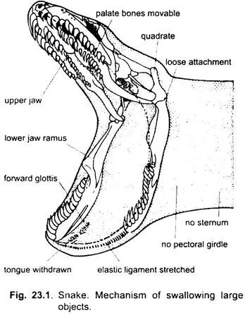
Snakes have long, narrow bodies, devoid of limbs. Moreover, some snakes have traces of hindlimbs placed at the root of the tail and carry a conical claw projecting at the sides of anus. The mouth and pharynx is extraordinarily distensible for swallowing their prey larger than the mouth.
The rami of the mandible are connected together only by elastic fibres at the symphysis, so that they can be widely separated. The maxillae, palatines and pterygoids are freely movable. Sternum and episternum are absent. As in lizards, a lachrymal duct carries away the tears and opens into the mouth in close relationship with the duct of Jacobson’s organ.
ADVERTISEMENTS:
Tympanum and tympanic cavity are absent. Eyelids are replaced by transparent spectacles developed from the eyelid rudiments. Skin is scaly which vary much in form, number and arrangement. When they are small and overlap, they are called scales, but when they are large and only touch by their edges, they are termed as shields. Snakes moult (ecdysis) several times in the course of the year. They strip off the whole of the scaly epidermis.
Vertebrae are very numerous and procoelous divided only into caudal and precaudal. Precaudal vertebrae except atlas carry ribs. Ribs articulate to the transverse processes of the trunk vertebrae and have only capitula. They are very movable in an antero-posterior direction. They are usually hollow and end ventrally in a cartilage attached to the connective tissue underlying the ventral shields.
Snakes run on the extreme points of their ribs which are moved forwards, carrying with them the ventral shields to which they are attached. Their tongue is long, narrow and forked, and retractile into a basal sheath. It is well provided with sense organs and is exceedingly protractile. It is used as a tactile organ. Majority of living snakes are non-poisonous.
Essay # 2. External Features of Snakes:
The body of snake is elongated, narrow and cylindrical, usually tapering towards the posterior end, and sometimes with or without a constriction behind the head. The short caudal region is indicated by the position of the cloacal opening. Forelimbs are never represented even by vestiges. But there are inconspicuous vestiges of hindlimbs in the form of small claw-like processes in pythons and other snakes.
ADVERTISEMENTS:
The mouth of a snake is capable of being very widely opened by the free articulation of the lower jaw. It mainly distinguishes it from the snake-like lizard. In snakes, the eyelids are immovable and the tympanum is absent.
Size:
Reticulated python (Python reticulatus) of India and Malaya (25 feet) and Anaconda (Eunectes murinus) of South America (25 feet and 300 lb) are the largest living snakes, Indo- Malayan King Cobra (Naja hannah) may grow up to 18 feet. Longest sea snakes grow to a length of 8 feet.
Locomotion:
Snakes move on land or in water by the movement of spinal column and muscles. This movement pushes the sinuous body horizontally against the resistant surrounding medium. Thick bodied snakes (e.g., boas, pythons, vipers) sometimes progress slowly in a straight line by means of contraction waves which pass along the costocutaneous musculature from head to tail. These specialised muscles are attached to the ventral scales which engage projections in the ground and so draw the animal along.
The North American side-winders (Crotalus) and Egyptian sand-vipers (Cerastes) move over sand by means of a series of horizontal double loops which engage the earth and propel the animal along sideways.
Various tropical and subtropical tree snakes glide across leaves and twigs from one tree to another.
The Asiatic Chrysopelea can ascend vertical walls and tree-trunks aided by laterally keeled ventral scales which engage very tiny projections.
Hearing:
ADVERTISEMENTS:
In snakes, tympanum, tympanic cavity and eustachean tube are absent. In spite of this, snakes appear to have a good sense of hearing. The columella auris occurs embedded in muscular and fibrous tissue and extends from the stapedial plate to the quadrate, against which it abuts by a cartilaginous epiphysis. In some snakes stapes is a bony plate enclosing the fenestra ovalis and without a shaft-like columella.
The snake is probably insensitive to air-borne sound. The rattle snake cannot hear its own rattling. The attachment of columella to quadrate undoubtedly makes the snake highly sensitive to earth-borne vibrations through quadrate, such as approaching feet. They are also quite sensitive to a narrow waveband of low frequency air-borne vibrations.
In snakes the tongue is long, forked at the tip and highly mobile and lodged in a sac in the floor of mouth. It is well provided with sense organs and used as a tactile organ. It can be protruded through an indentation at the extremity of the snout even when mouth is closed. Besides a tactile organ, the tongue of snakes presumably also acts as an auditory organ for receiving earth-borne vibrations.
Essay # 3. Feeding Mechanism of Snakes:
Snakes do not “slime” their victims before swallowing. Teeth are sharp-pointed, curved backwards and ankylosed to the jaws. They are present on the maxillae, palatines, pterygoids and on the dentaries. Teeth occasionally absent from pterygoid. They chiefly serve to hold the prey while it is being swallowed. Snakes feed exclusively on living animals, both warm- and cold-blooded. They swallow the prey without mastication.
Swallowing is effected thus- teeth on the lower jaw are alternately hooked further and further forwards into the body of prey (the two halves of the mandible moving forwards alternately), as a result of which mouth and pharynx of the snake are gradually drawn over the animal, the surface of which is at the same time made slippery by the secretion of the buccal glands.
During this process the larynx is projected forwards between the rami of the jaws, so that respiration can be maintained. After the completion of the laborious process of swallowing, the animal appears to be entirely prostrated and passes a long period in inactivity, during which slow digestion takes place.
Some snakes kill their prey by crushing, e.g., Python; some by poison and others, the majority, swallow their prey directly. Digestion begins in the stomach and is rapid. When a large animal is being slowly engulfed, the first part of the prey is partly digested before the hind-parts have been swallowed. After taking in a large meal, pythons are able to do without another for more than a year.
Besides pepsin secreted by fundic region of stomach, snake venom also assist in digestion. Swallowed rat by a boa constrictor, bones of its head have disappeared in two days and the whole skeleton in five days. Dasypeltis, the egg-eating snake, swallows the eggs whole and crushes them with special tooth-like processes of the neck vertebrae.
Essay # 4. Poison Apparatus in Snakes:
The poisonous snakes possess poison apparatus in their heads. It is absent in non-poisonous snakes.
The poison apparatus consists of a pair of:
1. Poison glands,
2. Their ducts,
3. A pair of fangs and muscles.
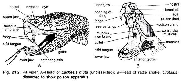
The poison glands are situated one on either side of the upper jaw. Each poison gland is sac-like (oval) in sea snakes or large and tubular in vipers.
In Naja naja, the poison gland is about the shape and size of an almond kernel. The gland is thickly encapsulated with fibrous connective tissue. It is composed of a body and a neck. The roughly fan-shaped capito-mandibularis superficial muscle, originating on the post-frontal (post-orbital) bone and parietal ridges, embraces much of the body of the poison gland.
Its contraction during biting, swiftly and synchronously squeezes poison into the poison duct. The duct passes forward along the side of the upper jaw and loops over itself just in front of the fang and opens either at the base of the fang or at the base of the tunnel on the fang.
1. Poison Glands:
The labial glands are present in a row in the upper and lower jaw. The posterior labial gland of the upper jaw is modified as the poison gland in the poisonous snakes. It is larger than the rest and different in structure.
2. Poison Ducts:
From the anterior end or neck of each poison gland arises a poison duct.
3. Fangs:
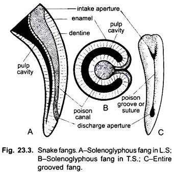
In poisonous snakes some of the maxillary teeth become poison fangs. These are usually larger than the ordinary teeth and are either grooved or perforated by a canal for the passage of the duct of the poison gland. Fangs are long, curved and pointed. The fangs regenerate by one of the small reserve fangs at its base (vipers). On the basis of structure and position, three types of fangs occur in poisonous snakes.
These are as follows:
(a) Solenoglyphous (solen = pipe + glyph = hollowed):
In Viperidae (vipers and rattle snakes), there is a single, large, curved, poison fang on the maxilla with small reserve-fangs at its base. These are the only teeth borne by the maxilla, which is very short. The large poison fang is capable of being rotated through a considerable angle, and moved nearly horizontal position (in which it lies along the roof of the mouth embedded in folds of the mucous membrane) to a nearly vertical position, when snake opens its mouth to strike its prey. Each poison fang contains a canal open at each end.
(b) Opisthoglyphous (opistho = behind):
In some poisonous snakes of the family Colubridae (subfamily Homalopsinae, Dipsadomorphinae and Elachiostodontinae) such as Dryophis, Elapops, Lycognathus, Elachistodon westermanni found in Bengal, etc., one or more of the posterior maxillary teeth grooved along its posterior border. There are more or less poisonous and poison is weak.
(c) Proteroglyphous (protero = first):
In sea snakes, Bungarus (krait), cobras (Naja), fangs (anterior maxillary teeth) are small, permanently erect and grooved along its anterior face.
(d) Aglyphous:
Fangs are lacking, so such snakes are non-poisonous.
4. Muscles:
The poison apparatus is associated with specialised bands of three types of muscles, viz.
(i) Digastric
(ii) Sphenopterygoid or protractor-pterygoid and
(iii) Anterior and posterior temporalis.
Digastric muscle is attached to the squamosal of the skull at one end and the articular of the lower jaw.
Sphenopterygoid is attached anteriorly to the sphenoidal region and posteriorly to the dorsal surface of the pterygoid. It helps in pulling the pterygoid forward Anterior and posterior temporalis muscles are attached to the sidewalls of the cranium and the lower jaw. They help in closing the lower jaw.
Essay # 5. Biting Mechanism of Snakes:
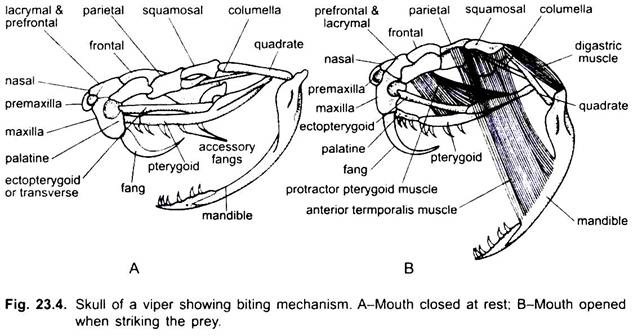
The skull and jaw bones in poisonous snakes are loosely and movably articulated, thus, allowing an enormous gape and swallowing whole of large prey. In cobras fangs are small and remain permanently erect, but in vipers the fangs are large and curved and lie against the root of mouth cavity when closed. Premaxilla, usually toothless and the bones of the upper jaw are loosely attached to rest of the skull. Quadrate jointed to the squamosal.
There are movable joints between the frontals behind and prefrontals and nasals in front and also between several other bones of brain case, palate and jaws. These joints have loose ligaments and allow movement in several directions and so permit a huge gap. The two halves of the lower jaw are connected together by elastic ligamentous tissue. So they are capable of being widely separated from one another.
The mechanism of biting is a complicated process and it can be described in the following four steps:
(i) Opening of the Mouth:
By the contraction of digastric muscles the mouth is opened (lower jaw moves down).
(ii) Rotation of Maxilla:
As the mouth opens the lower jaw moves down and the lower end of quadrate moves forward. Quadrate and squamosal are very movable. The pterygoid is movably attached to the palatine. Quadrate pushes the pterygoid forward and the pterygo-palatine joint bent.
This forward movement of the pterygoid is conveyed by the transpalatine bone to the maxilla and causes it to rotate through about 90° upon its prefrontal articulation in such a way that the surface to which the fang is attached is carried forwards and ventralwards, and the fang is erected, i.e., is made to project downwards at the front end of the mouth. The contraction of sphenopterygoid muscles also helps in the movement of pterygoid forward.
(iii) Closing of Mouth:
The closing of the mouth is brought about by the contraction of the temporalis and sphenopterygoid muscles. The point of fang is directed backward while the mouth is closed. It takes longer time to open the mouth than to close it.
(iv) Transference of Venom:
During the contraction of the digastric muscle the posterior ligament is relaxed and during the rotation of the squamosal bone the fan-shaped ligaments are stretched to squeeze the wall of the poison gland. This makes the poison to come out of the poison gland through the poison duct and the fang.
Essay # 6. Swallowing Mechanism of Snakes:
The rami of the lower jaw are separate in front but are connected by an elastic ligament which allows them to come apart. The upper jaw is loosely attached to the skull. A long quadrate articulates movably with the squamosal forming a lever which, when straightened, separates the lower jaw from the base of the skull, thus, the mouth and throat are opened so wide that the snake can swallow very large animals. Swallowing is done very slowly, and to enable breathing, the glottis is pushed forwards between the two rami of the lower jaw. Absence of pectoral girdle and sternum is an aid to swallowing large animals.
Non-Poisonous Snakes:
Fangs are not found. Teeth rarely developed on the premaxillae in snakes. But teeth are present on the maxillae, palatines and pterygoids, as well as the dentary of the mandible. Teeth are solid, elongated, sharp-pointed and usually strongly recurved. The function of such teeth is to hold prey and prevent it from struggling free while it is being swallowed.
Essay # 7. Snake Venom:
Snake venom is secreted by poison glands which are the modified supralabial or parotid glands present in the head of snakes. Venom is injected in the body of prey at the time of bite through the grooved maxillary fangs. Snake venoms vary in composition, most are mixtures of enzymes and proteins.
They often include polypeptides that poison the respiratory muscles of the prey. Many include proteases, which help in digestion. Venom is a clear sticky fluid of very light yellow or greenish colour. It is acidic in reaction and tasteless and odourless.
It causes no harm if swallowed but there should be no scratch in the interior of alimentary canal. It is only fatal when mixed with blood. It is soluble in water, glycerine or salt solution and in this condition also it is poisonous. It can be precipitated in silver nitrate and potassium permanganate.
Types of Venom:
The actual poison of venom appears to consist of albuminous substances in solution. It has different action in different snakes. Venom may (i) act on nerve cells and cause respiratory paralysis; (ii) destroy the endothelium of smaller blood vessels and allow blood to seep into the tissues; (iii) destroy erythrocytes and (iv) cause blood coagulation.
1. Neurotoxic Venom:
It causes the paralysis of the respiratory muscles. Venom of King Cobra (Naja hannah) and Australian Death Adder (Acanthophis) causes death due to respiratory paralysis. Venom of Tiger snake (Notechis scutatus) primarily affects the nervous system, although it is a powerful coagulant as well. Venom of Kraits is also neurotoxic. In India most deaths occur due to bite of Cobras, Kraits and Russels’ viper (Vipera russelli) whose venom is haemotoxic.
2. Haemotoxic Venom:
It is chiefly of vipers. It causes haemorrhage. The haemolysis and haemorrhage caused by the venom of South American Fer-de-lance (Bothrops atrox = lanceolatus) is so pronounced that bleeding occurs from the eyes, renal and alimentary tract epithelium.
Chance of survival from snake bite depends to a great degree on the amount of venom that snake habitually discharges, on its fang length and the depth to which they are driven into the tissues. It has been suggested that about 2 milligrams of neurotoxic venom of Tiger snake and Blue krait of India (Bungarus candidus) are toxic to the man.
Treatment:
The best treatment is to inject the antivenom serum or antivenin (serum of animals which have been rendered partially immune by repeated doses of the venom). Failing this, the best plan is to apply a ligature above the wound, twisting the string with a stick, and then to make an incision of the wound.
Then bandage the limb downwards towards the wound repeating this several times. Direct application into the widened wound of bleaching powder (calcium hypochlorite) or of a 1 per cent solution of Condy’s fluid is good. Amputation is of course the best remedy, if a very deadly snake has bitten the part. Alcohol and injection of ammonia are useless, and sucking the wound is dangerous. Many mammals are said to be immune, e.g., mongoose, hedgehog and pig.
Essay # 8. Non-Poisonous Snakes:
1. Typhlops:
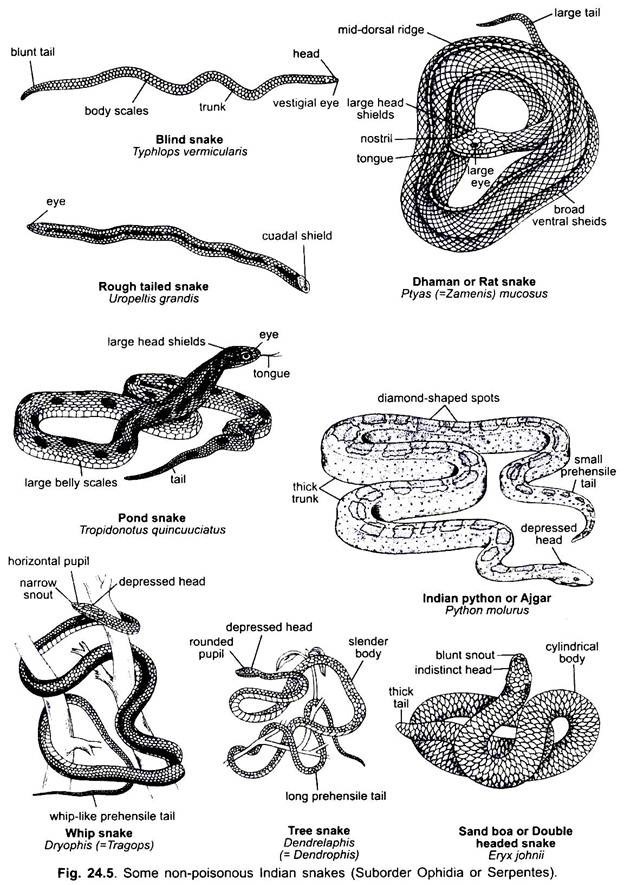
Typhlops (Fig. 24.5) is commonly known as blind snake. It has a large number of species in various tropical and marshy regions of the earth. It lives underground and looks like an earthworm. It has a brown cylindrical body generally 25 cm long, but various species range from 17.5 cm to 60 cm in length. The body is covered uniformly with smooth, rounded, overlapping scales.
Head is small and non-expansible. There is no distinct neck. Eyes are covered by scales so that it is blind. Tail is short and stumpy. It differs from other snakes in having a few teeth on the upper jaw only. Squamosal and transverse bones of the skull are absent. Quadrate is fixed, rami of the lower jaw are joined, and mouth is non-expansible. It has vestiges of the pelvic girdle. It is insectivorous and oviparous.
2. Python:
Python (Fig. 24.5) is a large, massive, non-poisonous snake covered with small scales and shields on the head and large plates below. Tail is prehensile. It has a vertical pupil. Premaxilla also bears teeth. It constricts warm-blooded animals in its coils and kills them due to suffocation, then it slowly swallows the prey. Pythons and Boas have vestigial pelvic girdle which articulates with a small femur bearing a claw. The claws are seen on either side of the cloaca, they play a part in copulation.
They are oviparous. Python molurus, the Indian python, is over 8 metres long. It is grayish-brown with red and black spots and a mark like a spearhead between the eyes, ventral side is yellowish. Scales of upper lip have pits. It is nocturnal and very often waits on trees for its prey. It loves to lie in water. Python reticulatus of Myanmar, Indo-China and Malaya is 10 metres long and is the longest known snake. Pythons are found in Asia and Africa.
3. Eryx:
Eryx johnii (Fig. 24.5) is commonly called Indian or John Sand Boa or Dumuhi. It is a uropeltid, found in dry, hot, sandy parts of India, Africa and Sri Lanka. The body is short and flat with a stumpy tail, eyes are minute. The head is pointed and resembles the tail, hence, it is often called a two-head boa.
It has a uniformly sandy brown colour, it may have cross bars and dark spots, with small smooth scales above and slightly enlarged plates below. Eryx is 1 metre long. It is a harmless, sub-terranean snake. It lives in burrows of small rodents on which it feeds, it also feeds on lizards and worms.
4. Zamenis (Ptyas):
Zamenis mucosus (Fig. 24.5) is commonly called dhaman or rat- snake of India. It is a highly active non-poisonous snake which feeds on rats, small rodents, and frogs. It grows to a length of 2.5 metres, and has an elongated tail. Scales above are smooth with pits, below they are large transverse bands. Colour is brownish above with black bands on the tail in some, it is pale yellow ventrally.
The eyes are large with round pupil. It has a sharp ridge along the backbone. Ptyas is very swift, ill-tempered and bites viciously. It has a nervous temperament and is untamable. It is oviparous. It is regarded as a friend of the farmers because it destroys rats. It is found in India, Myanmar and Java.
Essay # 9. Poisonous Snakes:
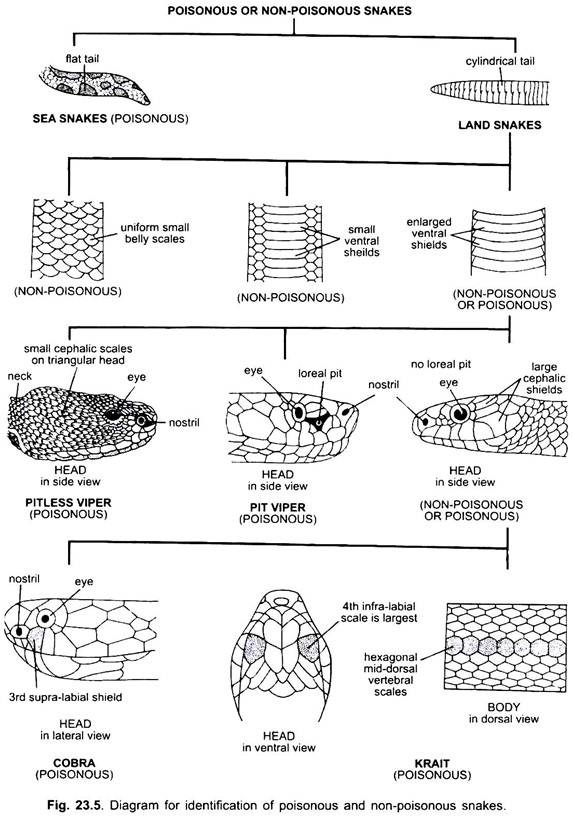
The poisonous snakes are kraits, cobras, pitless vipers and pit vipers, sea snakes, coral snakes and rattle snakes. These possess poison glands and fangs-modified maxillary teeth. Their tails except sea snakes are cylindrical. Heads are covered either with small scales (vipers and sea snakes) or with large shields. Ventrals cover the entire width of the belly so that laterals are not visible from the ventral side.
1. Bungarus:
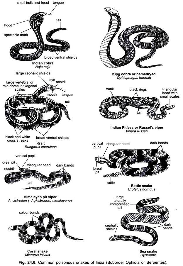
Bungarus (Fig. 24.6) is commonly called krait. It has several species in India and Malaya. It is slender and about 1 metre long, of pale brown colour with darker bluish-black markings having white cross bars, ventral surface is white. Fangs are small. Scales are smooth. The backbone is ridged having a central row of enlarged scales. It is oviparous and the female incubates the eggs.
It feeds on toads, mice and smaller snakes. The common krait, Bungarus caeruleus, comes into homes, is slow moving, but is highly poisonous. Its venom is more poisonous than that of a cobra. Bungarus fasciatus is the banded krait about 1.20 metres long. It has black and yellow subequal cross bars and the back is keeled. It is found in Indonesia and India.
2. Naja:
Naja (Fig. 24.6) is commonly called Indian Cobra or nag. Naja naja is also called Naja tripudians. It is a highly poisonous snake growing up to 2 metres in length, of brown or blackish colour. It lives in holes and under stones, but also comes into human dwellings. It feeds on frogs, lizards, rats and other snakes. Scales are smooth, dorsal surface of the head has small scales and large shields.
The head is small. The cervical ribs when raised form a hood on the sides of the neck, its hood spread is more than in other cobras. Pupil is round. Fangs are small and relatively non-movable. On each side is a large poison gland enclosed in a fibrous capsule, its duct runs into a canal in the fang. When annoyed, it raises the front part of the body about 30 cm, spreads its hood and hisses through the nose.
It bites with a chewing motion making several wounds. The venom is neurotoxic and fatal. It is more deadly than that of vipers. There are three common varieties of cobras in India according to the makings on the hood. On the ventral side all have two black and white marks. 1. But on the dorsal side of the hood the diocellate cobra has a black and white spectacle mark. 2. The monocellate cobra has a single black ring enclosing a pale area having a black spot in the centre. 3. The black cobra has no mark but the scales are barred. The cobra is oviparous and the female incubates the eggs. Cobra is found also in Malaya, Iran, Myanmar, and southern China.
Naja hannah (king cobra also called Hamadryad), is found in India, Malaya, and southern China. It grows up to 6 metres, but has a diameter of only 7.5 cm, the average length is 3.5 metres. It is the world’s largest poisonous snake and the most deadly and venomous of all snakes.
The venom injected is large in quantity and causes quick death due to respiratory paralysis. The king cobra has a small head, the hood spread is only about 10 cm, on the hood are transverse bands and white marks.
The colour is variable, it has a pale gray, fawn, olive, or black colour with dark cross bands which appear like rings, the end of the tail may be yellowish. It is oviparous and feeds on non-poisonous snakes, carefully avoiding poisonous ones. The king cobra is very intelligent, dangerous in habits, and bad-tempered, it is bold and often attacks without provocation.
3. Vipera:
Vipera (viper) (Fig. 24.6) has thick body up to 4.3 cm long. Head is large, flat, and covered with small scales. Nostrils are lateral, eyes are far forwards and there is a sensory pit between the eye and the nostril in some. Pits are sensory organs with which the snake can detect its prey. The scales on the body are keeled. Tail is short and tapers abruptly.
The Indian Russel’s viper, Vipera russelli, has enlarged nostrils unlike other vipers. It is pale brown above with three longitudinal series of black rings, each ring is bordered with white, ventrally it is yellowish. Scales on the head are small and keeled. In vipers, the maxilla is small and bears long, movable fangs with canals. The fangs are erected for biting and folded back against the roof of the mouth when not in use.
Behind the fangs there are no teeth in the upper jaw, but there are two rows of teeth on the palatines. Vipers are nocturnal. In biting it opens its mouth very wide and strikes like lightning, thrusting its long fangs, the poison is deadly. Vipers are viviparous and are found in India, Myanmar and Sri Lanka, other vipers have species in Europe, Africa and Asia.
4. Ancistrodon or Agkistrodon:
Ancistrodon himalayanus (Fig. 24.6) is commonly called Himalayan pit viper. The general colour is bluish-brown above with dark brown or black spots appearing like cross bars and the ventral side is brown with black and white spots. Body is not much elongated, measuring about 60 cm in length. The scales covering the body have apical sensory pits.
Mouth is grey with black spots. The upper lip is slightly raised in the rostral region. Supra-labials are 5-7, 3rd and 4th are enlarged but not touching the eye. Post-oculars are two, the last greatly enlarged and pre-oculars are three. A loreal pit is present in between the eye and the nostril on each side. Loreal pits function as thermoreceptors. Each pit has two cavities separated by a membrane.
These organs (and the pits in the marginal scales along the mouth of pythons and some boas) are supplied by branches of the trigeminal nerve. This apparatus perhaps helps the animals to locate precisely and attack warm-blooded prey. Eyes are big with golden iris and vertical pupils. Tail is cylindrical without rattles. Viviparous.
Poisonous and its poison is haemotoxic. Ancistrodon is quite a timid snake, remains hidden under stones, etc. Ancistrodon himalayanus is distributed in hilly regions of Eastern parts of India, Sri Lanka and Java. It is very common in Kashmir and is found even at an altitude of about 3658 metres.
5. Hydrophis:
Hydrophis (Fig. 24.6) is commonly called Sea snake. The body is long and laterally compressed posteriorly. Head and neck are very slender. The general colour is dark olive green above with yellowish cross bars and white below. The ventral scales are small. Tail is laterally compressed and acts like paddle in swimming. Eyes are small with rounded pupil.
Loreal shield is absent. One pre-ocular, two post-oculars and 7-8 supra-labials of which 3rd and 4th touching the eye. Presence of 14-18 maxillary teeth behind the poison fangs. Viviparous. Carnivorous, feeding on fishes, etc. Hydrophis is deadly poisonous and its venom is neurotoxic. Hydrophis occurs in the Bay of Bengal and Malay Archipelago.
6. Crotalus:
Crotalus (Fig. 24.6) is commonly called rattle snake. It has several species in America and Mexico. It grows up to 2.5 metres and has a prominent sensory pit between the eye and nostril. Upper side of head has small scales. Fangs are long and movable, they are folded back when not in use.
At the end of the tail is a rattle having a small hard button at the tip and usually 10 to 12 dry horny rings, the rings are formed as hollow segments by the end of the moulted skin being left attached to the tail. The rattle is always held up vertically. It makes a grating sound in locomotion and is probably for warning. The rattle snake is highly poisonous. It is carnivorous and viviparous.
