In this article we will discuss about:- 1. Meaning of Antibody 2. Distribution and Production of Antibody 3. Basic Structure 4. Classification of Molecules.
Meaning of Antibody:
The term antibody refers to glycoproteins synthesised in response to the administration of an antigen and which react specifically with that antigen and to a variable extent, with molecules of similar structure. From the classical experiment by A. Tiselius and E. A. Kibat (1939), it became evident that the antibodies reside in a particular serum protein fraction — the Ƴ-globulin fraction.
Since then, antibodies contained in Ƴ-globulin fraction were called immunoglobulins, to distinguish them from any other proteins that might be contained in the Ƴ-globulin fraction. Advances in electrophoresis technology now shown that some classes of antibody molecules are also present in the β and α fractions of serum in addition to Ƴ fraction.
Distribution and Production of Antibody:
In vertebrate bodies, antibodies are distributed in fluids like plasma, mucosal secretions and in interstitial fluid of tissues. They are found on the surface of a limited number of cell types. Within B lymphocytes, antibodies are present in cytoplasmic membrane bound compartments (endoplasmic reticulum, and Golgi complex) and on the surface, where these molecules are expressed as integral membrane proteins.
ADVERTISEMENTS:
Secreted form of antibodies is present in body fluids as mentioned above. Antibodies synthesised and secreted by B cells often attach to the surface of certain other immune effector cells, such as mononuclear phagocytes, NK (natural killer) cells and mast cells which have specific receptors for binding the antibody molecules.
In blood, antibodies remain in serum. Serum that contains a detectable number of antibody molecule that is capable of binding to a particular antigen is called antiserum. A healthy adult human produces about 3gm of antibodies everyday most of which belong to IgA class.
Antibodies that enter the circulation have limited half-lives, for example IgG, the most common type of antibody found in serum has a half-life of about 3 weeks.
Basic Structure of Antibody:
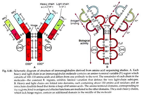
All antibody molecules are glycoprotein in nature and have a common structure of four polypeptide chains; two identical light (L) chains and two identical heavy (H) chains. Each light chain is about 24 kD and each heavy chain is of 55 to 70 kD.
ADVERTISEMENTS:
Each light chain is bound to one heavy chain by a strong disulfide bond. The two light chains and two heavy chains are also linked to each other by disulfide bridges forming the basic four-chain immunoglobulin structure, the dimer (HL)2.
The first 110 or so amino acids of the amino terminal region of a light or heavy chain varies greatly among antibodies of different specificity. These segments of highly variable sequence are called V regions: VL in light chain and VH in heavy chain (Fig. 6.46). The regions of relatively constant sequence beyond the variable region are referred to as C regions: CL in the light and CH in the heavy chain.
Both light and heavy chain variable regions participate in antigen recognition. The carboxyl terminal constant regions of the heavy chains mediate effector functions. The immunoglobulins are glycoproteins; the sites of attachment of carbohydrates are restricted to the constant regions. The carbohydrates increase the solubility of immunoglobulins and thus help in their clearance from serum by liver.
ADVERTISEMENTS:
Amino acid sequencing of Light and Heavy chains:
Amino acid sequencing of light chain indicated that there are two light chain types, kappa (ƙ) and lambda (λ). In human 60% of the light chains are kappa and 40% are lambda types. A single antibody molecule contains only one light chain type, either ƙ or λ chain but never both. The amino acid sequences of λ chains show minor differences. In human there are four subtypes of λ chain and in mice there are three subtypes.
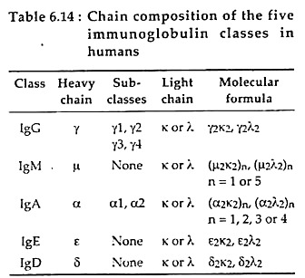
Heavy chain sequencing revealed the presence of five types of chains, viz., µ, δ, Ƴ, ɛ and α. Each of these five different heavy chains is called iso=-type. The heavy chain of a given antibody molecule determines the class of that antibody (see classification of antibody). Each class can have either ƙ or λ light chain.
A single antibody molecule has two identical heavy chains and two identical light chains, H2L2, or a multiple (H2L2)n of this basic four-chain structure (Table 6.14). Differences in the amino acid sequences of a and y chain led to further classification of human a chain into α1, α2 and Ƴ chain into four subclasses (Ƴ1, Ƴ2, Ƴ3 and Ƴ4).
Fine Structure of Immunoglobulins:
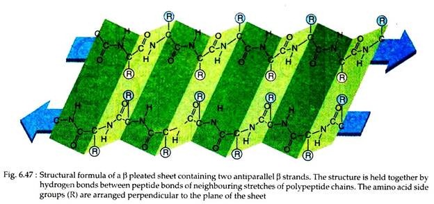
The primary, secondary, tertiary and quaternary organisations of the proteins determine the structure of different immunoglobulin molecules. The amino acid sequence is the primary structure, which accounts for the variable constant regions of both chains. The extended polypeptide chain folds back and forth upon itself into an antiparallel β pleated sheet.
Thus, the secondary structure is formed. The chain then again folds into a tertiary structure of compact globular domains, that remain connected to neighbouring domains by continuation of polypeptide chain lying outside the β pleated sheet.
Finally, the adjacent heavy and light chains interact through their globular domain to form the functional domain or quaternary structure that enables the antibody to specifically find antigen (Fig. 6.46B).
ADVERTISEMENTS:
Careful analysis showed that the light chains contain one variable (VL) and one constant (CL) domain, heavy chains contain one variable (VH) and either three or four constant domains (CH1, CH2, CH3 and CH4), depending on their type. Each domain is made of about 110 amino acids both in light and heavy chain.
Immunoglobulin fold:
In X-ray crystallographic analysis, immunoglobulin domains appear to be folded into a characteristic compact structure called immunoglobulin fold. In such folds two P pleated sheets remain in a ‘sandwich’ form, each containing anti-parallel β strands of amino acids. The β strands within a sheet are stabilized by hydrogen bonds connecting-NH group in one strand with -CO group of the adjacent strand (Fig. 6.47).
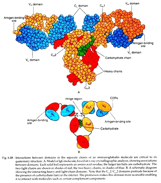
The β strands contain alternately hydrophobic and hydrophilic amino acids whose side chains remain arranged perpendicular to the plane of the sheet. The basic structure of the immunoglobulin fold contributes to the quaternary structure of immunoglobulin by facilitating non-covalent interactions between domains across the faces of the P sheets (Fig. 6.48A, B).
The structure of the folds also allows for -variable lengths and sequences of amino acids that form the loops connecting the β strands. Some of such loops of VH and VL domains constitute the antigen binding site of the molecules.
Structure of Variable Region Domains and their Relationship to Antigen Binding:
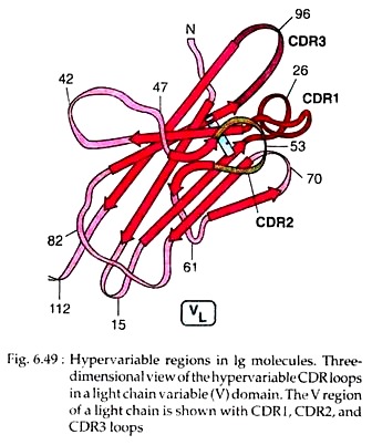
The differences in the specificity among antibodies are actually due to the differences in the amino acid sequence of particular areas within the variable regions on both light and heavy chains. These areas are called hyper-variable regions (HV).
Three such hyper-variable regions are present in human heavy and light chain, constituting 15%-20% of the variable domain, each about 10 amino acid residues long. The remainder of the VL and VH domain, exhibit far less variation; these stretches are called the framework regions (FRS).
In an antibody molecule, the three HV regions of a VL domain and the three HV regions of a VH domain are brought together in three-dimensional space to form an antigen-binding surface. Because these sequences form a surface that is complementary to the three-dimensional structure of the bound antigen, the hyper-variable regions are also called complementarity determining regions (CDRs) (Fig. 6.49).
Proceeding from either the VL or the VH amino terminus, these regions are called CDRl, CDR2 and CDR3, respectively. The CDR3s of both the VH segment and VL segment are the most variable of the CDRs. Crystallographic analysis of antibodies reveals that the CDRs form extended loops that are exposed on the surface of the antibody and are thus available to interact with antigen (Fig. 6.49).
In general, more residues in the heavy-chain CDRs appear to contact antigen than in the light- chain CDRs. Thus the VH domain often contributes more to antigen binding than the VL domain. Such dominant role of heavy chain in antigen binding has been proved by a study.
In that study, a single heavy chain specific for a glycoprotein antigen of human HIV was mixed with various light chains of different antigenic specificity. Such hybrid antibodies were then allowed to bind to the HIV glycoprotein antigen.
A high degree of antibody-antigen binding indicated that the heavy chain alone was sufficient to confer specificity as the light chains were nonspecific. However, in some antibody-antigen reactions, the light chain makes the more important contribution.
Advanced X-ray crystallographic analyses have revealed that in some cases binding of antigen induces conformational changes in the antibody, antigen or both. For example, binding of neuraminidase and anti-neuraminidase is accompanied by a change in the orientation of side chains of both the epitope and the antigen binding site.
Actually, such conformational changes lead to closer fit between the epitope and the antibody’s binding site. Another striking example of such conformational change has been seen during binding of monoclonal antibody against HIV protease and the peptide epitope of the protease. In this case, upon binding of the antigen, the CDR1 region of the light chain moves as much as 0.1 nm and CDR3 of heavy chain moves 0.27 nm.
Such conformational changes due to movements of CDRs enable the antibodies to assume a shape more effectively complementary to that of their epitopes. In this binding, conformational changes were not limited to the antibody; the enzyme’s native conformation also undergoes some changes resulting in the firm binding of protease enzyme with anti-HIV protease.
Constant region domains:
The constant region domains of immunoglobulin molecules perform various biological functions:
i. The CH1 and CL domains can extend the Fab arms of the antibody molecule and can cause maximum rotation of the Fab arms. Thus facilitating the interaction with antigens.
ii. The constant region domains hold the variable domains of both heavy and light chains by virtue of the inter-chain disulfide bond between them.
iii. The CH1 and CL domains also contribute to antibody diversity by allowing more random associations between VH and VL domains than would occur if this association were driven by the VH/VL interaction alone.
iv. The above two domains may also contribute to the overall diversity of antibody molecules that can be expressed by an animal, since these two can increase the number of stable VH and VL interactions.
Hinge regions:
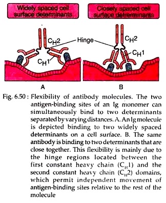
The IgG, IgD and IgA isotypes of immunoglobulin contain a proline rich residues between the CH1 and CH2 domains of their Ƴ, δ and α chains, respectively. This region is called hinge region. This hinge region is highly flexible and varies from 10 to more than 60 amino acids residues in different isotypes.
Portions of this sequence assume a random and flexible conformation, permitting molecular motion between CH1 and CH2. Some of the greatest differences between the constant regions of the IgG subclasses are concentrated in the hinge. This leads to different overall shapes of the IgG subtypes.
The number of inter-chain disulfide bonds in the hinge region varies considerably among different classes of antibodies and between species. The and e chains of IgM and IgE isotypes lack hinge region, but they have an additional domain of 110 amino acids (CH2/CH2) that has hinge-like features.
The large number of proline residues in the hinge region gives it an extended polypeptide conformation, making it particularly vulnerable to cleavage by proteolytic enzymes particularly with papain or pepsin. Another amino acid cysteine is also rich is hinge region and it forms inter-chain disulfide bonds that hold the two heavy chains together.
It was revealed by X-ray crystallographic study, that the hinge region serves as a sort of tether that allows the Fab segment and the Fc to move relative to each other (Fig. 6.50). In this way, the Fab arms can move and twist to align the CDRs with epitope displayed on cell surface.
The Fc arm can move to maximise various affecter functions such as those of the complement system or bind to cell surface receptors specific for the Fc region.
Reducing antibody structure:
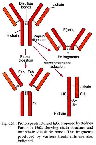
Most information of antibody structure as described above came from the experimental studies done by Rodney Porter and Gerald Edelman (1960s). In one experiment, Porter subjected IgG to brief digestion with the enzyme papain and separated the fragments.
Brief papain digestion cleaves only the most susceptible bonds of IgG and produces two identical fragments, each with a molecular weight of 45kD: one called Fab fragments (“fragment, antigen binding”) because they had antigen binding activity, and the other fragment (mol. wt. of 50kD) called the Fc fragment (“fragment, crystallizable”), because it was found to crystallize during cold storage (Fig. 6.51).
A similar experimental approach, but with the enzyme pepsin, was performed by Alfred Nisonoff. Such digestion of IgG molecule produced one single fragment (mol. wt. of 100kD) composed of two Fab-like fragments. This F(ab’)2 fragment unlike the Fab fragments was able to precipitate antigens. However, pepsin digestion never produced Fc fragment, because it had been digested into multiple fragments (Fig. 6.51).
Porter also confirmed that the IgG molecule of 150kD molecular weight was composed of two 50kD polypeptide chains, designated as heavy (H) chains and two 25kD chains, designated as light (L) chains. The relation of enzyme digestion products Fab, F(ab’)2 and Fc to the heavy chain and light chain reduction products was also experimentally confirmed by Porter.
In his experiments with goat antisera, he found that antibody to the Fab fragment could react with both the H and L chains, whereas antibody to the Fc fragment reacted only with the H chain.
These observations led him to conclude that Fab consists of portions of a heavy and a light chain and that Fc contains only heavy chain components. Thus, from different experimental observations, Poter and Edelman proposed the IgG structure according to what this antibody molecule consists of two identical H chains and two identical L chains, which are linked together by disulfide bridges.
Structural differences between secreted immunoglobulin (slg) and membrane-bound immunoglobulin (mlg):
Antibodies may be expressed in secreted or membrane-bound forms, which differ in the amino acid sequence of the carboxyl terminal end of the heavy chain C regions. In the secreted form of antibody molecules as found in blood and other extracellular fluids, the sequence of the last CH region terminates with charged and hydrophilic amino acid residues.
But, in the membrane bound antibody, as found only on the plasma membrane of B-cells, the carboxyl-terminal domain contains three regions:
i. An extracellular hydrophilic “spacer” sequence composed of 26 amino acid residues.
ii. A hydrophobic trans-membrane sequence.
iii. A short cytoplasmic tail.
In mlgM and mlgD molecules, the cytoplasmic tail of heavy chain is short, only 3 amino acid residues in length, while in mlgG and mlgE molecules, it is somewhat longer, up to 30 amino acid residues in length. The immature B cells, called a pre-B cell, express only mlgM. mlgD appears later in maturation and is co-expressed with IgM on the surface of mature B cells before they have been triggered by antigen.
A memory B cell can express mlgM, mlgG, mlgA or mlgE. Even when different classes are expressed sequentially on a single cell, the antigenic specificity of all the membrane antibody molecules is identical, so that each antibody molecule binds to the same epitope.
Secreted IgG, IgE and all membrane Ig molecules regardless of iso-type, are monomeric with respect to the basic antibody structural unit (i.e., they all contain two heavy chains and two light chains). In contrast, the secreted forms of IgM and IgA form multimeric complexes in which two or more of the four-chain core antibody structural units are covalently joined.
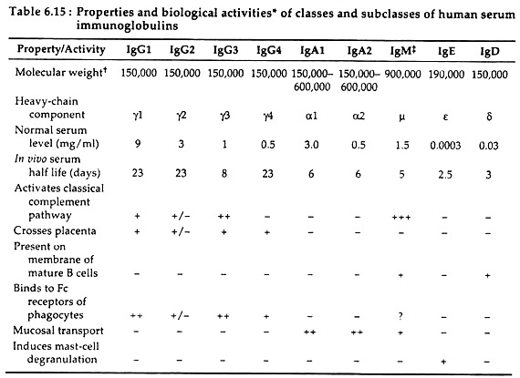
These complexes are formed by interactions between regions, called tailpieces that are located at the carboxyl terminal of p and a heavy chain respectively (Table 6.15). Multimeric IgM and IgA molecules also contain an additional 15-kD polypeptide called the joining (J) chain. It is disulfide bonded to the tail pieces and serve to stabilise the multimeric complexes.
Classification of Antibody/Immunoglobulin Molecules:
Antibody or immunoglobulin molecules can be divided into distinct classes and subclasses on the basis of differences in the structure of their heavy chain C regions. These are also called isotypes and subtypes respectively. Different isotypes and subtypes of antibodies perform different effector functions.
1. Immunoglobulin G (IgG):
TSG is the major serum immunoglobulin making up 80% of total serum immunoglobulin.
Structure:
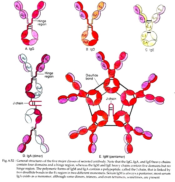
The IgG molecule consists of two heavy chains (Ƴ) and two k or two λ light chains. Its molecular weight is 150kD (two L chains of 22,000 Daltons each and two H chain of 55,000 Daltons each). As an IgG has got two identical antigen binding sites, it is called divalent (Fig. 6.52A).
Subtypes:
In human, the heavy chain exists in four different forms and there are four subtypes of IgG viz., IgG1, IgG2, IgG3 and IgG4 (see Table 6.15). The amino acid sequences that distinguish the four IgG subclasses are encoded by different germ-line CH genes, whose DNA sequences are 90%- 99% homologous. These subclasses also show differences in their hinge regions and in the number and position of inter-chain disulfide bonds.
Secretion and existence:
IgG appears later in serum about 2 weeks after infection, but persists for long duration. IgG has a longest half-life of about 23 days. It is the predominant antibody in secondary immune response and its production requires T cell help.
Its catabolic rate depends directly upon the total IgG concentration in serum. When IgG level in serum is raised, as in myeloma, kala-azar, the synthesised IgG against a particular antigen, is catabolized rapidly.
Functions:
Except IgG2, other readily cross the placenta and protect the developing foetus.
i. IgG3 is a most effective complement activator followed by IgG1. IgG2 is less efficient while IgG4 is not able to activate complement at all.
ii. IgG1 and IgG3 bind with a high affinity to Fc receptors on phagocytic cells and thus mediate opsonisation.
iii. IgG coats microbes and targets them for phagocytosis by neutrophils and macrophages.
Besides these specific functions, IgG also helps in antibody dependent cell: mediated cytotoxicity, feedback inhibition of B cells etc.
2. Immunoglobulin A (IgA):
IgA constitutes only 10%-15% of the total serum immunoglobulin, but it is the predominant antibody in external secretions like breast milk, saliva, sweat, tears, mucus of bronchial, genitourinary and digestive tracts.
Structure:
In serum, IgA exists primarily as a monomar (MW 160kD), but polymeric forms such as dimers, trimmers, and even tetramers, all of which contain a J-chain polypeptide are sometimes seen. The IgA of external secretions is called Secretory IgA. It consists of a dimer or tetramer, a J-chain polypeptide, and a polypeptide chain called secretory component (Fig. 6.52D).
The J-chain helps in polymerisation of both serum IgA and secretory IgA. The secretory component is a 70kD polypeptide, produced by the epithelial cells of mucous membranes. It consists of five immunoglobulin-like domains that bind to Fc region domain of the IgA dimer by disulfide bonds.
Subtypes:
Some IgA exists as monomer (H2L2). Two subclasses of IgA are known, IgA1 and IgA2. The former is dominant (60%) form in the secretion and constitutes only a minor component of serum IgA. IgA2 lacks inter-chain disulfide bonds between H and L chains.
Secretion and existence:
IgA is diminished within the epithelial cells of glands, intestine and respiratory tract with J chain. This dimeric IgA then combines with another protein, the secretory component; ultimately the complex is transported across the cytoplasm and secreted into body fluids.
Everyday a human secretes from 5 gm. to 15 gm. of secretory IgA into mucous secretions. Its production requires T cell help and special mucosal stimulation to promote production of slgA. It has got a half-life of 6 days.
Functions:
Secreted IgA acts as an important effector molecule at mucous surface, the main entry sites of most pathogens. Binding of slgA to bacterial (e.g., Salmonella, Vibrio cholerae) and viral (e.g., Polio, Influenza etc.) surface epitopes prevent viral infection and bacterial colonisation by their adherence to mucosal cells.
The complexes are then trapped by mucous and are eliminated by ciliated epithelial cells of respiratory tract or by peristalsis of the gut.
i. IgA in breast milk may protect the newborn against infection during the first few months of life.
ii. IgA does not fix complement, but it is capable of activating the alternate complement pathway.
iii. It is also an effective opsonin, reacting with FcαR on monocytes and neutrophils.
3. Immunoglobulin M (IgM):
IgM is the largest of all immunoglobulin molecules in terms of size and thus is essentially restricted to vascular fluid. It accounts for 5%-10% of the total serum immunoglobulin. It is the first immunoglobulin class produced in a primary response to an antigen and to be synthesised by the neonate.
Structure:
IgM is secreted by plasma cells as a pentamer in which five monomer units are held together by disulfide bonds that link their carboxyl-terminal heavy chain domains (Cµ4/Cµ4) and link their Cµ3/Cµ3 domains (Fig. 6.52E). The five monomer subunits are arranged with their Fc regions in the centre of the pentamer and the ten antigen-binding sites on the periphery of the molecule.
Each pentamer contains an additional Fc-linked polypeptide chain; J chain which is disulfide bonded to carboxyl terminals of two of the ten p chains. The J chain is added just before secretion of the pentamer. The theoretical valency of IgM is 10 which is observed on interactions with small haptens only. With larger antigens the effective valency falls to 5, probably due to the lack of flexibility in the molecule.
Secretion and existence:
The appearance of IgM on blood precedes IgG in phylogeny. As it is the earliest synthesised immunoglobulin in foetus, it appears in blood in about 20 weeks of age. Some antibodies, such as anti-A, anti-B antibodies to typhoid O antigen, rheumatoid factor and W. R. antibodies in syphilis belong to IgM class. IgM remains largely confined to blood stream (80%) because of its large size it cannot spread from blood into the tissue.
It has got a half-life of 5 days and it can be produced in a T- independent manner.
Functions:
Because of high valency, IgM is more efficient than other Ig isotypes in binding multidimensional antigens like virus and red blood cells.
i. As it has two Fc regions in close proximity in the pentameric structure of a single molecule, IgM is more efficient than IgG in activating complement.
ii. It is also a good agglutinator for bacteria and blood protozoan.
iii. IgM also plays an important accessory role as a secretory immunoglobulin.
iv. IgM is particularly effective for immunity against polysaccharide antigens present on the exterior of pathogenic bacteria.
4. Immunoglobulin E (IgE):
IgE constitutes less than 0.1% of the total immunoglobulin. It mediates immediate hypersensitivity reactions that are responsible for the symptoms of hay fever, asthma, hives and anaphylactic shock.
Structure:
Structurally IgE resembles IgG molecules. It has molecular weight of 200kD (see Fig. 6.52C).
Secretion and existence:
It is mainly produced by a very small proportion of plasma cells living in the respiratory and intestinal tracts. It has got a short half-life of 2-3 days.
Functions:
During allergic reactions IgE mediates its activity. IgE bound to mast cells or basophils serves as a receptor for allergens and parasitic antigens. Cross linkage of receptor-bound IgE molecules by antigen induces degranulation of basophils and mast cells, as a result, a variety of pharmacologically active mediators in the granules are released, giving rise to allergic manifestations.
i. IgE plays an important role for protection against parasitic infection.
ii. It is responsible for anaphylactic hypersensitivity.
5. Immunoglobulin D (IgD):
This class of antibody was detected in myeloma protein and constitutes only about 0.2% of total immunoglobulin in serum. It is least well-characterised among immunoglobulins from functional aspect.
Structure:
Structurally it resembles IgG molecule. Molecular weight of IgD antibody is 150kD. It has a monomeric structure (Fig. 6.52B).
Secretion and existence:
IgD remains present together with IgM on the membrane of mature B cells.
Functions:
IgD antibody is thought to function in the activation of B cells by antigen. No biological effector function has been clearly identified for IgD.
