The central nervous system is composed of the brain and the spinal cord. Impulses are transmitted to the central nervous system through afferent (sensory) fibres of the peripheral nerves. The brain interpretes the messages and despatch them to the effectors through efferent (motor) neurons.
Brain:
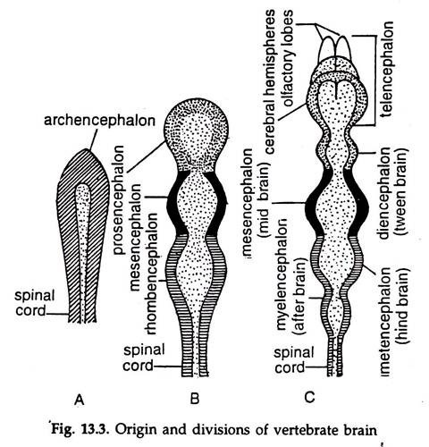
In the embryo, the brain is formed by the dilation of the neural tube, at the anterior end. Constrictions appear in the brain rudiment and three vesicles, the forebrain, or pros-encephalon, midbrain or mesencephalon and hindbrain or rhomb encephalon are formed. The forebrain subdivides into an anterior telencephalon and a posterior diencephalon.
The midbrain remains unchanged as mesencephalon. The hindbrain gives rise to an anterior metencephalon (cerebellum) and a posterior myelencephalon or after brain, the medulla oblongata (Fig. 13.3). The rest of the neural tube form the spinal cord in the adult.
The neurocoel or the cavity in the neural tube divides correspondingly forming ventricle in the brain. The chief divisions of the ventricle are the first, third and fourth ventricles located in the telencephalon, diencephalon and medulla oblongata, respectively.
Paired lobes, the cerebral hemispheres develop from the pros-encephalon and their cavities are the first and second ventricles, also known as lateral ventricles, communicate with the third ventricle in the diencephalon by a narrow foramen of monro.
The aqueduct of Sylvia’s joins the third ventricle with the fourth ventricle in the metencephalon and myelencephalon. The fourth ventricle is continuous with the central canal (neurocoel), which extends up to the end of the spinal cord.
The canal is lined by a ciliated epithelium. The ventricles and the central canal are filled with a cerebrospinal fluid. The solid portion of the central nervous system enclosing ventricles in the brain and central neural canal in the spinal cord is made of grey and white matter.
ADVERTISEMENTS:
Grey matter:
Aggregation of the cell bodies of the neurons gives a grey appearance to the region formed by them and the portion is called grey matter.
White matter:
The Portion formed by the aggregation of nerve processes is whitish in colour and termed white matter. In brain, the grey matter is restricted to the outer zone, while in the spinal cord it lies inwards, towards the central canal and vice versa.
ADVERTISEMENTS:
The brain and the spinal cord are en-sheathed in meninges, consists of three layers. The outermost layer, Dura mater lies against the cranium bone, the innermost layer, the pia mater lies on the surface of the brain and the middle layer, the arachnoid lies in between the two. The spaces between the pia mater and the arachnoid are filled with cerebrospinal fluid.
Modifications of vertebrate brain:
Starting from distinct five divisions which is found in fishes, the vertebrate brain undergoes a series of modifications resulting in the most complex brain in mammals. The primitive brain becomes complicated due to unequal thickening of the walls.
The floor of the medulla thickens greatly but the roof is made of a single layer of nortnervous epithelium. The great thickness of the cerebellum obliterates the epicoele. The ventral wall of the mesencephalon thickens to form two longitudinal bands, the crura cerebri; the dorsal wall forming a pair of large optic lobes.
The sides of the diencephalon thickens to give rise paired optic thalami, the roof remaining a single layer thin membrane, but a part of it forms pineal apparatus. The floor of the diencephalon grows downward to form infundibulum to which the pituitary gland is attached.
The pituitary gland is formed partly from a diverticulum of the pharynx and partly from the extremity of the infundibulum. The telencephalon grows forward, enlarges greatly and due to fusion of the internal dorsal and ventral edges of the lateral walls, the two cerebral hemispheres (cerebrum) are formed.
With the progress of evolution, the horizontal plain of the vertebrate brain underwent drastic changes. Owing to the bending down of the anterior part (cerebral flexure) the brain assumed the shape of a retort, the axis of the forebrain inclined to that of the hind brain. The cerebral hemispheres grew forward, parallel to the hindbrain. The thickening of the floor of the mid and hindbrain made them obscured in the adult.
The grey column of the spinal cord extend up to the medulla. In the brain, the column breaks up into aggregated cell bodies, the nuclei. The dorsal nuclei are sensory nuclei and contain cell bodies of afferent neurons from the cranial nerves. The ventral nuclei are motor nuclei, as they contain the cell bodies of efferent neurons.
Cerebral hemispheres:
ADVERTISEMENTS:
The olfactory lobes are associated with the anterior region of the cerebral hemispheres. The cortex of the cerebral hemispheres known as cerebral cortex exhibits zonal specialisation to carry specialised functions.
These may be placed in three groups:
a. Memory, intelligence, thinking, reasoning, sense of responsibility and moral sense, word perception and writing in man.
b. Sensory perception. Touch, sight, hearing, taste, smell, temperature, pain, etc.
c. Voluntary muscle initiation and control.
Mesencephalon:
Groups of nerve cells and nerve fibres in the mesencephalon connect the cerebrum (cerebral hemispheres) with the posterior part of the brain and the spinal cord.
The nerve cells function as relay centres for ascending (afferent) and descending (efferent) nerve fibres:
Cerebellum:
The cerebellum is concerned with coordination of voluntary muscles, posture, movement, balance and equilibrium of the body.
Medulla oblongata:
In the deeper structure lie groups of cells associated with reflex activity. The medulla acts as cardiac centre, respiratory centre, vasomotor centre and reflex centre.
Reticular formation:
An aggregation of neurons in the core of the brain stem is known as reticular formation. It is surrounded by neural pathways conducting impulses between the brain and the spinal cord.
Numerous synapses connect it with other parts of the brain and enable it to constantly receive both sensory and motor impulses. The reticular formation is considered to be a primitive part of the brain. Reticular formation is concerned with consciousness or awareness of the environment.
Spinal cord:
The spinal cord is a hollow, cylindrical tube, suspended in the neural canal of the vertebral column and surrounded by meninges and cerebrospinal fluid. It runs backward from the medulla oblongata and extends from the upper border of atlas to very close to the posterior of the vertebral column (lower border of the first lumber vertebra in man).
Two types of tissues constitute the spinal cord. The grey matter surrounding the central canal is X or X-shaped in transverse section. Nerve cell nuclei and synapses are present in the grey matter. The white matter surrounds the grey matter.
Myelinated nerve fibres running longitudinally and so connecting different levels of the spinal cord are present in the white matter. The spinal cord can carry out certain functions, e.g., spinal reflexes, independent of the brain. Extensive connections between sensory and motor nerves in the spinal cord enable it to carry such activities.
