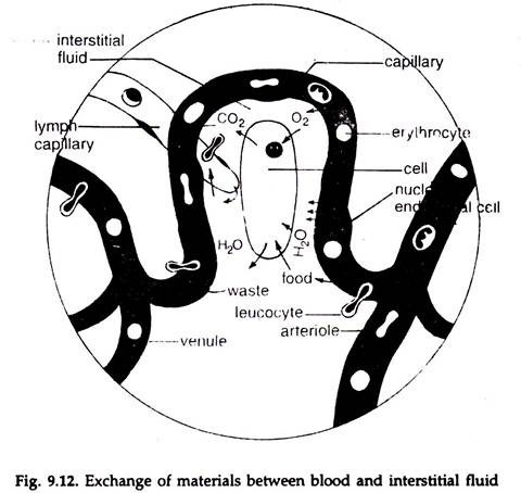
In this article we will discuss about the capillary exchange of materials between blood and interstitial fluid in animals.
Blood is composed of a liquid component, the plasma, blood cells, protein and other materials. About 55 per cent of the blood volume is plasma. The plasma contains about 90 per cent water; free in solution are 7 to 8 per cent soluble proteins, 1 per cent electrolytes and 1 to 2 per cent other materials, viz. glucose, amino acids, lipids, vitamins, hormones, metabolic intermediates and nitrogenous wastes.
The blood in the arteries moving to the whole of the body, except respiratory organs, is rich in oxygen and low in carbon dioxide. The blood in the ventral aorta of fishes and pulmonary arteries of land and secondarily adapted aquatic vertebrates, moving, to the respiratory organs, is low in oxygen and rich in carbon dioxide.
On its way an artery branches to a number of arterioles which have a diameter of 0.1 mm or less and an arteriole subdivides into many capillaries, the average diameter being 8-10 µm; the total cross-sectional area increases, which causes a gradual fall of velocity of blood flow at each step. The total cross-sectional area of all the capillaries of an arteriole is greater than that of the arteriole and the result is decreased velocity of blood flow.
ADVERTISEMENTS:
Further, the friction of the moving blood with the blood vessels causes continuous fall of mean blood pressure. The fall of pressure continues in the capillaries and veins. The velocity of blood flow begins to increase with the flow of blood into the venule. Several venules join to form a vein, several veins to a larger vein, and at each step the cross- sectional area decreases, leading to faster flow of blood.
In animals with open circulatory system the haemolymph flows freely into sinuses from the arterioles. Cells are bathed in haemolymph and exchange of materials takes place across the plasma membrane of the cells.
In animals with closed circulatory system, the blood flows into the capillaries. The capillaries are numerous closely packed, extremely thin-walled, narrow tubes with a diameter 8.0 to 10.0 µm. The capillary walls are formed by membranes of the endothelium and bear numerous minute pores, the diameter of which may be quite large, as 100 nm in some glands.
These openings are closed only by delicate protein fibres constituting the basement membrane. In addition, at many points the endothelial cells are not firmly united with one another and they overlap, leaving direct passages through the wall.
ADVERTISEMENTS:
Following the concentration gradient, water and dissolved substances with small molecular weight pass from the capillaries to the interstitial fluid, vice versa, and from interstitial fluid to cells through plasma membrane and reverse.
Oxygen, glucose, amino acids, vitamins, ions, etc. diffuse out through the capillary walls to the interstitial fluid and from there move to the cells across the plasma membrane. Carbon dioxide, nitrogenous wastes and other products of cell metabolism move out to the interstitial fluid through plasma membrane and from there to the capillaries through capillary walls (Fig. 9.12).
Water moves each way through the capillary walls.
Several forces help in maintaining a constant pressure of interstitial fluid. Most protein molecules are large and those with molecular weight of about 70,000 or more cannot pass through the capillary wall.
The pressure of plasma in the capillaries, the hydrostatic pressure or blood pressure, which varies along the length of the capillaries, causes water and dissolved substances to filter out of the capillaries, while proteins remain in the capillaries and exerts an osmotic pressure, colloid osmotic pressure, also called protein osmotic pressure (protein’s hunger for water), which is fairly constant along the length of the capillaries. The higher hydrostatic pressure at the arterial end of the capillaries gradually decreases towards the venous end with the flowing out of water.
In the exchange process, opposite forces are at work. At the arterial end the hydrostatic pressure is greater (35 mm) in the beginning of capillary than the protein osmotic pressure (25 mm Hg) and there is a net force, the filtration pressure, that forces out water and dissolved materials, including some plasma proteins out of the capillaries.
With the fall of pressure in the capillaries, less fluid is filtered out and when the hydrostatic pressure is less than the colloid osmotic pressure, the process is reversed.
The outflow of water causes higher protein osmotic pressure at the venous end, which is higher than the hydrostatic pressure and there is a net force, the absorption pressure that draws water and dissolved materials into the capillaries.
In most cases, the filtration pressure is a bit higher than the absorption pressure. Absorption surface of the capillaries at the venous end is slightly more and more water can move in through it.
The amount of fluid exchange through the capillary wall fluctuates. Usually the outflow exceeds the inflow and the excess interstitial fluid passes into lymph capillaries and a constant pressure is maintained in the cell environment.
The interstitial fluid entering the lymph capillaries is known as lymph. The lymph capillaries join to form lymph ducts, that drain into larger ducts, which finally open into the venous system and the fluid is returned to the blood.
