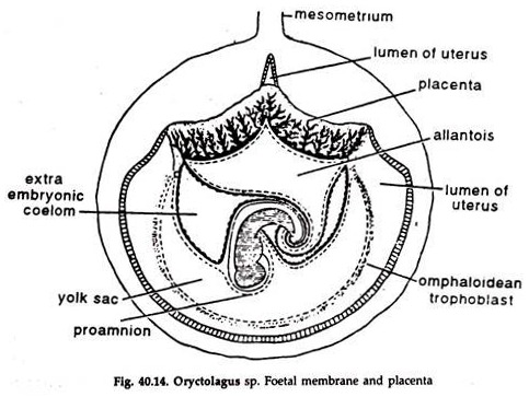
In this article we will discuss about the formation of placenta in oryctolagus, explained with the help of a suitable diagram.
Changes in the Embryo:
1. After fertilization the zygote descends in the oviduct, undergoes cleavage, and a blastocyst, corresponding to the blastula of lower forms is produced.
2. The epiblast formed during gastrulation is differentiated into an inner cell mass and a sub-zonal layer, the trophoblast.
ADVERTISEMENTS:
3. The trophoblast is rapidly differentiated into an outer layer—the plasmotrophoblast, and an inner layer or layers of cytotrophoblast.
Implantation of the Ovum:
1. A pair of folds arise upon the mesometric side of the uterus and the blastocyst lies in between the folds and becomes attached by the trophoblast.
2. The enzymic activity of the trophoblast erodes the uterine epithelium, and the embryo becomes surrounded by a cushion of liquefied cells and blood called histotroph.
3. The embryo sends finger-like projections or villi in the uterine tissue.
Appearance of Primary Villi:
ADVERTISEMENTS:
1. The plasmotrophoblast soon turns to a solid mass of multinucleated and vacuolated protoplasm.
2. The vacuoles are termed primary lacunae.
3. The plasmotrophoblast sends projections into the uterine mucosa and pushing out the somatic cells approaching the capillaries, establishes communication between the lacunae and the capillaries.
4. The lacunae break tip the trophoblast into trabeculae giving the entire chorion a villus appearance, the primary villi.
Secondary or True Chorionic Villi:
ADVERTISEMENTS:
The cytotrophoblast also forms villi communicating with the chorion and these are called true villi.
Later development:
1. In the beginning, the chorionic villi surround the whole embryo but later all except those between the uterine wall and the chorion atrophy.
2. The region where the chorionic villi are restricted is known as chorion frondosum.
3. The placenta now consists of the villi of the chorion frondosum and part of the uterus, in which the villi are lost.
Nutrition in Placenta:
1. Histotrophic nutrition. Nutrition is supplied from the mother to the embryo.
2. Haemotrophic nutrition. Trophoblast penetrates the trophospongia facilitating the osmotic interchange of fluid and diffusion of gases between the foetal and maternal blood.
Functions of Placenta:
1. Anchorage of the foetus to the uterine wall.
2. Protection of the foetus.
ADVERTISEMENTS:
3. Supply of nutrition to the foetus from maternal blood through the umbilical cord.
4. Storehouse of fat and glycogen.
5. Acts as a foetal lung and a foetal kidney.
6. Acts as a barrier against bacterial infection.
