In this article we will discuss about the process of gametogenesis and fertilization in animals, explained with the help of suitable diagrams.
Spermatogenesis:
1. It occurs in the testes. The walls of the seminiferous tubules of vertebrates contain primordial germ cells.
2. The primordial germ cells begin to enlarge with increased metabolism and undergo mitotic division, and produce spermatogonia.
ADVERTISEMENTS:
3. The spermatogonium divides meiotically (first meiosis) resulting in two spermatocytes.
4. Due to second meiotic division (mitosis) of spermatocyte four haploid spermatids are formed.
5. Spermatids metamorphose to spermatozoa. In the process spermatid increases in size; centriole divides into two; distal centriole forms the main axis of the tail; the axial filament is surrounded by a fibre coat; mitochondria form a spiral sheath; Golgi complex form acroblast and give rise to acrosome.
6. The mature spermatozoon has three distinct zones—head, trunk and tail. It can move actively in fluid.
ADVERTISEMENTS:
Structure of Human Spermatozoa:
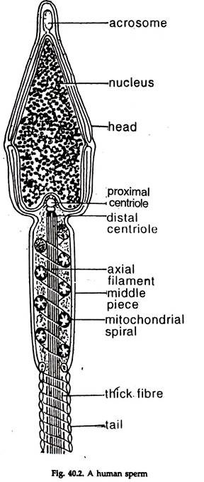
In human, the spermatozoon (Fig. 40.2) measures 60 µm long and 2.5 to 3.5 µm in diameter at the broadest point in the head. The spermatozoon is rich in deoxyribonucleic acid (DNA).
Head:
ADVERTISEMENTS:
The head is constituted by an anterior acrosome (galea capitis) and a nucleus.
a. Acrosome:
Acrosome is a double-walled, granulated sac, convex anteriorly and flat posteriorly. It contains enzymes to dissolve the zona pellucida (egg membrane) of the ovum to facilitate entry of the sperm for fertilization.
b. Nucleus:
It is a dense structure, rich in DNA, narrower anteriorly and bears a depression at the middle of the broad posterior end.
Middle piece:
It connects the head with the tail piece. The proximal centriole of the anterior end fits into the depression of the head and the distal centriole is attached with the nine axial filaments in the mid piece. Elongated mitochondria form a firmly coiled spiral around the axial filaments. The middle piece provides energy for locomotion of sperms.
Tail piece:
It has two portions:
ADVERTISEMENTS:
a. The broad anterior main piece and
b. A narrow, posterior terminal end piece.
a. The main piece of the tail consists of a spiral mitochondrial sheath surrounding a group of eleven fibrils. Two fibrils are centrally placed and the rest form a ring around them. Contraction and relaxation of peripheral fibrils help the spermatozoa to move forward. The enzyme adenosine triphosphatase (ATPase) is present in the whole length of the main piece.
b. Terminal end piece (flagella). It is the slender, last portion of the tail piece. The fibrils are naked and resemble cilia.
Oogenesis:
1. It occurs in the ovary. The phases of division in multiplication are similar to those in spermatogenesis.
2. The primary germ cells of the ovary undergo several mitotic divisions to produce oogonia.
3. The oogonium gives rise to primary oocytes mitotically.
4. A primary oocyte undergoes first meiotic division and produces one large secondary oocytes and one small first polar body or polocyte.
5. The oocyte and the polocyte divide mitotically (second meiotic division). The oocyte produces one large ootid and one small second polar body and the polocyte two polar bodies.
6. In oogenesis one haploid ootid and three polar bodies are formed. On maturity, the ootid is transformed into ovum. The ova are of one type only. The nucleus with haploid chromosome number is termed pronuclear.
Structure of Ova:
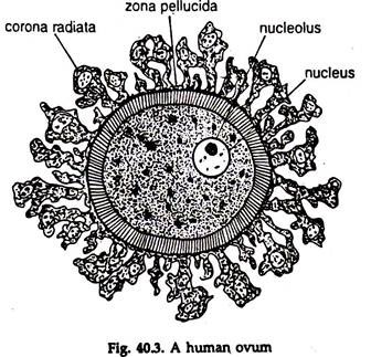
The ovum is a large, round structure. In human, according to the stage of development, it measures 117 to 142 µm in diameter. The cytoplasm of the ovum is termed yolk or ooplasm, the nucleus as the germinal vesicle and the nucleolus as germinal spot.
The yolk comprises:
(a) Cytoplasm similar to that of other cells and
(b) Deutoplasm or nutritive yolk, consists of fatty droplets containing lecithin, a phospholipid.
The mode of distribution of deutoplasm in the ovum differ in different animals. During oogenesis mitochondria increase in numbers and become uniformly distributed in the cytoplasm. The Golgi apparatus spreads out, being restricted usually near the periphery. The amount of yolk in the ovum varies in different groups of animals.
The mature ovum or egg is surrounded by two membranes, the plasma membrane (egg membrane) and the vitelline membrane. The vitelline membrane is thin and surrounds the plasma membrane. In addition to these primary membranes, secondary membrane secreted by the ovary and tertiary membrane secreted by the glands in the oviduct may surround the egg.
In branchiostoma, only the plasma membrane and vitelline membrane are present. In frogs and toads a sphere of jelly around the egg is enclosed in a thin membrane. In birds, a two-layered shell membrane and a calcareous shell are secreted by the oviduct and, in mammals, the additional layers— zona pellucida and chorona radiata are possibly primary and secondary membrane, respectively, (Fig. 40.3).
Types of Eggs:
The eggs are classified on the amount and position of deutoplasm (yolk) present in them.
Homolecithal or Isolecithal egg:
The amount of yolk is small and present chiefly in droplets and minute spherules, uniformly distributed in the cytoplasm. True homolecithal eggs are found in eutherian mammals.
Microlecithal egg:
The amount of yolk is small.
Examples: Cnidarians.
Megalecithal egg:
The amount of yolk is large.
Examples: Some invertebrates, reptiles, birds and prototherian mammals.
Centrolecithal egg:
The yolk is present around the nucleus in the form of a large sphere.
Example: Arthropod eggs.
Telolecithal egg:
Eggs are large, megalecithal and the yolk occupies almost the entire egg, except a minute area in the animal pole. In chordates, telolecithality occurs in varying degrees.
a. In Branchiostoma, the yolk is concentrated at one pole, the region of future endoderm cells.
b. In cyclostomes and amphibians, the quite high amount of yolk is concentrated in the vegetal pole.
c. In reptiles and birds, the amount of yolk is large and occupies almost the entire egg and the active cytoplasm. The egg nucleus forms a small cap on the yolk in the animal pole.
Fertilization:
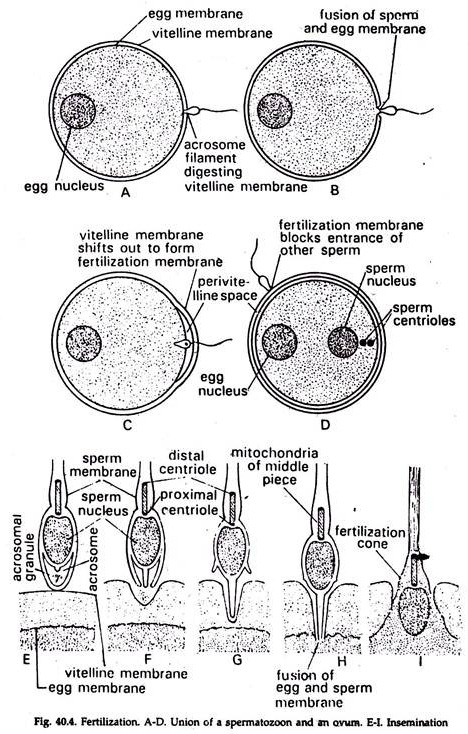
Fertilization is a complete fusion of sperm and ovum except the chromosomes, resulting in a single diploid cell, the zygote. The union of the nuclei of male and female gametes is karyogamy and that of their cytoplasm is plasmogamy.
The steps in fertilization (Fig. 40.4) are:
I. Approach of the spermatozoon to the egg:
a. By chemotactic movement the sperm swims towards the egg.
b. The jelly coat of the egg produces fertilizin and the sperms produce antifertilizin.
c. The fertilizin and the antifertilizin combine to form an initial bond to facilitate penetration of spermatozoon into the egg.
II. Penetration of the sperm:
a. The bound or agglutinated spermatozoon produces an enzyme, lysine, which dissolves egg membranes in the local area and clears the path for spermatozoon to reach the egg surface.
b. The acrosomal filament is pushed through the jelly and vitelline membrane, and touches the surface membrane of the egg cytoplasm.
III. Reaction in egg during sperm penetration:
a. Coming in contact with the acrosomal filament the egg cytoplasm bulges forward to produce a conical projection, the fertilization cone.
b. It gradually engulfs the spermatozoon and carries it inward.
c. Simultaneously profound cortical reaction occurs in the cytoplasm.
d. Immediately after penetration of the spermatozoon, the vitelline membrane separates from the plasma membrane. The vitelline membrane thickens and forms the fertilization membrane which prevents entry of other sperms and thus ensures entrance of only one sperm into the egg.
e. The cortical granules swell, become liquefied and the fluid is released in the space between egg cytoplasm and fertilization membrane.
IV. Behaviour of sperm within the ovum:
a. The head and middle piece enter the egg cytoplasm but the tail is left outside.
b. The egg completes its meiosis immediately and loses the centriole.
c. The sperm rotates 180° and its middle part comes to the front end. The path along which the sperm moves within the egg is known as fertilization path.
d. The egg pro-nucleus moves to the periphery and the movement of the sperm is oriented to it. This is copulation path.
e. The sperm pro-nucleus swells up. The centrosome and the centriole in the middle piece form aster around the two pronuclei.
f. The sperm centriole divides into two, move to the poles, being connected by fibres.
g. The nuclear membranes disappear, the chromosomes of the male and female pronuclei are arranged in the equator of the spindle.
h. It is similar to metaphase plate of meiosis and immediately followed by the cleavage of the zygote.
Importance of Fertilization:
a. The sperm entry activates the secondary oocyte to complete its maturation division.
b. Restores diploid number of chromosomes and recombines the maternal and paternal genetic materials.
c. Evokes development and cleavage of egg to form a complete organism.


