In this article we will discuss about the mechanism of gas exchange in various animals:- 1. Aquatic Animals 2. Invertebrate Gill 3. Vertebrate Gill.
Aquatic Animals:
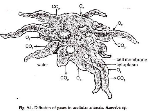
Small aquatic animals, protozoa and somewhat large forms, viz. sponges and hydras, in which water current bathes the cells, a definite respiratory structure is absent. The situation is similar in forms in which the body surface acts as the respiratory membrane (Fig. 9.1).
The oxygen dissolved in water directly moves into the cells through plasma membrane and carbon dioxide diffuses out. In stagnant water, organelles like cilia develop to maintain a water current, which brings oxygen supply and remove carbon dioxide from the surface.
In larger invertebrates, specialised respiratory structures, ctenidia have developed. The ctenidia may be located in different regions of the body in different animals, but, as a rule, they are restricted to the anterior part of the body, in the majority. The ctenidia are present in association with Para podia in annelids, on both sides of the cephalothorax in crustaceans and in the mantle cavity in molluscs.
Invertebrate Gill:
ADVERTISEMENTS:
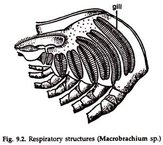
A ctenidium (gill) is made of a number of lamellae. Each lamella is composed of a large number of gill filaments. The lamellae are attached to a central axis. Channels in the central axis carry blood to and from spaces in the lamellae. Gas exchange occurs in the gill filaments. In crustacea, the ctenidia (Fig. 9.2) on the sides of the cephalothorax, lie in the branchial chambers formed by the sides of the carapace.
In prawn the chambers communicate with the exterior along their anterior, posterior and ventral borders. Movements of the scaphognathites of the second maxillae set a constant backward to forward water current in the gill chambers.
ADVERTISEMENTS:
In crabs, the carapace is sealed along the ventral margin and the water enters through apertures at the bases of the great chela, the first pair of walking legs. In molluscs, the gills are lodged in the mantle cavity. In a bivalve, by the beating movement of cilia present in the gill filaments, a current of water enters the mantle cavity through the inhalant siphon and escapes to the exterior through exhalant siphon.
In prosobranchs, contraction of muscles in the mantle and nuchal lobes enlarges the mantle cavity and water enters through the left nuchal lobe or the siphon and goes out through the right siphon due to relaxation of muscles in the mantle.
Vertebrate Gill:
The respiratory organs of adult cyclostomes and fishes are internal gills. Some amphibians respire with larval gills during development, which takes place in water.
Structurally, the respiratory organs of cyclostomes and Chondrichthyes (Elasmo-branchii) exhibit some differences between them and also with Osteichthyes (Teleostomi), but in all cases, the gills are located in the lateral walls of the pharynx, hence called pharyngeal gills.
ADVERTISEMENTS:
During embryonic development a series of paired outgrowths appear in the lateral walls of the pharynx, extending from inside to outside. These are gill pouches. The out pushing’s or gill pouches finally open to the exterior by narrow slits, the gill slits or gill clefts.
Thus, each gill pouch is in communication with the pharyngeal cavity by an internal and with the outside by an external branchial aperture. Two successive gill pouches are separated by a fibrous partition (the original pharyngeal wall), the inter-branchial septum.
The membrane lining the anterior and posterior walls of each gill pouch is folded into a number of horizontal ridges, the branchial filaments. The filaments are richly supplied with blood, where gaseous exchange takes place.
The visceral arches supporting the pharynx are a series of U-shaped rods. The visceral bar or each half of the arch is lodged in the pharyngeal side of an inter-branchial septum, alternate with the gill pouches. As a result, a visceral arch bears the posterior set of filaments of one pouch and the anterior set of the filaments of the next one.
In Osteichthyes (Teleostomi) the inter-branchial septa are reduced to narrow bars enclosing the visceral arches of the pharyngeal wall and a double row of gill filaments develop from each visceral bar. A gill made of one set of gill filaments is hemi-branch or half gill and a gill with two hemibranchs is holobranch.
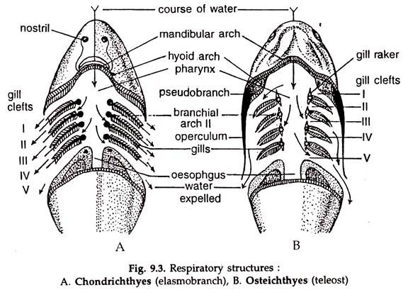
In cyclostomes and elasmobranchs there are several pairs of gill pouches (Fig. 9.3). A gill pouch is a biconvex sac, containing numerous highly vascular gill lamellae. The gill pouches open into the pharynx. In lampreys, several pairs of gill slits are present, while in hag fishes, the gill slits are only one pair. In all cases the gill slits are separated by partition. The number of paired gill slits vary in different Chondricthyes.
In teleost’s, the partition between two gill slits is reduced to a branchial arch bearing two rows of gill filaments (Fig. 9.3). All the gills of each side are placed in a gill chamber, covered externally by a bony operculum (L. operculum = lid).
ADVERTISEMENTS:
A thin branchiostegal membrane is present at the posterior margin of the operculum. During respiration the gill chamber becomes tightly closed by pressing the membrane against the body wall.
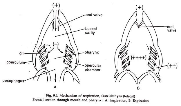
A respiratory current is established by lowering the floor of the buccal cavity. The water rushes in the cavity through the open mouth (Fig. 9.4A). With the closure of the mouth and the oesophagus and raising of the buccal floor and contraction of the pharyngeal walls, water enters the gill pouches and passes out through gill slits in cyclostomes and elasmobranch (Fig. 9.3A).
In teleost’s with raising of the buccal floor and contraction of the pharyngeal walls, the branchiostegal membranes are forced open and water from the buccal cavity is driven to the gill chamber through the gill slits and thence to the exterior (Fig. 9.3B, 9.4B).
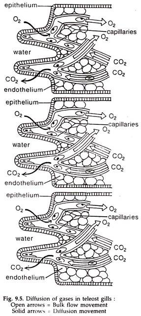
Afferent and efferent branchial arteries are lodged in the gill arch. The former carries deoxygenated blood to the gills and the latter drains away oxygenated blood from the gills (Fig. 9.5).
For an active respiration the gills must have a large surface area and a large volume of water must move across the gill lamellae or filaments. Oxygen is only slightly soluble in water and the weight of the water passing through the gill slits is about 0.1 million times the weight of available oxygen. Usually, water moves in one direction across the gills.
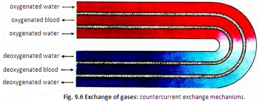
Diffusion of gases between the water and the blood or haemolymph in the gills is rather rapid. The diffusion is accelerated by a mechanism termed counter-current exchange in some crustaceans, molluscs and fishes.
In the process, the direction of water flow across the gills is opposite to the flow of blood or fluid (Fig. 9.6). About 90 per cent of dissolved oxygen may be removed from water by the counter- current exchange.
