The below mentioned article provides an overview on Fasciola Hepatica (Liver Fluke):- 1. Meaning of Fasciola Hepatica 2. Habit and Habitat of Fasciola Hepatica 3. Morphology 4. Digestive System 5. Nervous System 6. Reproductive System 7. Life History 8. Infection to Secondary Host (Snail) 9. Infection to New Primary Host (Sheep).
Contents:
- Meaning of Fasciola Hepatica
- Habit and Habitat of Fasciola Hepatica
- Morphology of Fasciola Hepatica
- Digestive System of Fasciola Hepatica
- Nervous System of Fasciola Hepatica
- Reproductive System of Fasciola Hepatica
- Life History of Fasciola Hepatica
- Infection to Secondary Host (Snail) of Fasciola Hepatica
- Infection to New Primary Host (Sheep) of Fasciola Hepatica
1. Meaning of Fasciola Hepatica:
Liver flukes are typical digenean trematodes and are commonly called “flatworms” or “flukes” on account of their flat, leaf-like structure. Fasciola hepatica is the common liver fluke of sheep. It is the first trematode whose life history was described by Thomas in 1883. It is of much importance as it causes fascioliasis—a disease that causes damage to liver- tissues and bile ducts of sheep.
ADVERTISEMENTS:
Etymology:
Latin: fasciola, small bandage
The species name hepatica stems from the Greek word hepar meaning liver.
2. Habit and Habitat of Fasciola Hepatica:
The sheep liver fluke is an endoparasite which completes its life history in two hosts. The adult flukes are typical parasites of vertebrate animals but one stage of their life history is invariably spent in an invertebrate host — a mollusc. Thus, they have a digenetic life history for which the group to which they belong has been named as Digenea.
ADVERTISEMENTS:
Sometimes the adult flukes invade other vertebrates like goat, horse, dog, ass, ox, rabbit, monkey and even man. They cause serious loss of human life and domestic animals. The disease caused by this parasite is known as liver rot. A single sheep may harbour as many as 200 liver flukes in its liver. F. hepatica has a cosmopolitan distribution and is common in areas where sheep and cattle are being reared.
3. Morphology of Fasciola Hepatica:
External Morphology:
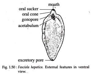
F. hepatica is a soft-bodied, flattened leaf-like animal and exhibits bilateral symmetry. They measure about 1 to 2.5 cm in length and about 1 cm in width. The anterior end of the body (Fig. 1.50) is drawn out into a prominent conical projection, termed the oral cone or head lobe, bearing at its tip a somewhat triangular aperture, the mouth and surrounding it is the oral or anterior sucker.
On the ventral surface, a little behind the head lobe, is situated a much bigger sucker called the ventral or posterior sucker (also known as acetabulum). Between the two suckers and close to the posterior sucker is situated the genital opening through which the penis sometimes protrudes. At the extreme posterior tip of the body lies the excretory aperture, which is a single opening.
ADVERTISEMENTS:
The canal of Laurer opens on the middle of the dorsal surface. The body surface is marked by the presence of a number of conical projections— the spinules or papillae which are extensions of the cuticle surrounding the body.
Internal Morphology:
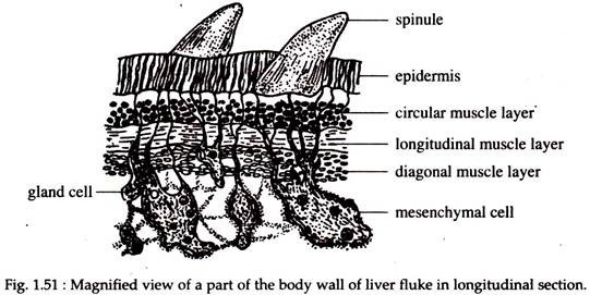
The architecture of the body wall of F. hepatica is peculiar and consists of the following succession of layers (Fig. 1.51):
(i) An outermost region, the epidermis (formerly called cuticle) from which spinules arises;
(ii) A basement membrane;
(iii) Circular, longitudinal and oblique or diagonal muscle fibres; and
(iv) A number of unicellular gland cells with protoplasmic processes. Interspaces between the organs are packed with parenchyma cells. Many ectodermal dells are seen to sink into the parenchyma and are connected to the cuticle by protoplasmic projections (Fig. 1.51).
4. Digestive System of Fasciola Hepatica:
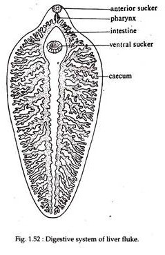
The mouth at the centre of the muscular oral sucker, leads to a small rounded bulblike pharynx which is muscular and suctorial (Fig. 1.52). This leads to a short passage, the oesophagus and is followed by the intestine. It divides immediately into two main limbs; — right and left and runs to the posterior side of the body.
ADVERTISEMENTS:
From each of these limbs are given off, both internally and externally, a number of blind branches or caeca. The caeca of the inner side are short and simple, while those of the outer side are longer and branched. Practically every region of the body is traversed by the caeca due to its ramification.
The intestine and its branches are very conspicuous as they remain filled with decomposed products of the blood on which the fluke feeds. As anus is absent there is no aperture of communication between the intestine and the exterior.
Excretory System:
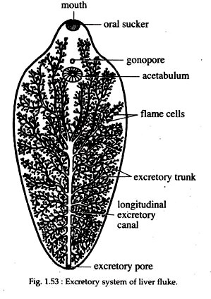
Excretory system is a branching system of vessels, the water vessels that ramify mouth throughout the body. It consists of a median longitudinal main trunk or pro-tone-phridial tubule which opens to the exterior by means of the excretory pore (Fig. 1.53) situated at the posterior end of the body.
From the anterior region of the main canal four large canals are given off, each of which branches repeatedly. These then give off smaller vessels and these again to still smaller twigs or capillaries which enter the inconspicuous flame cell or flame bulb or protonephridia.
The flame cells excrete water and waste metabolites, such as unwanted iron from haemoglobin. A curious factor in Fasciola is that its excretory canals normally contain an abundance of fat droplets.
5. Nervous System of Fasciola Hepatica:
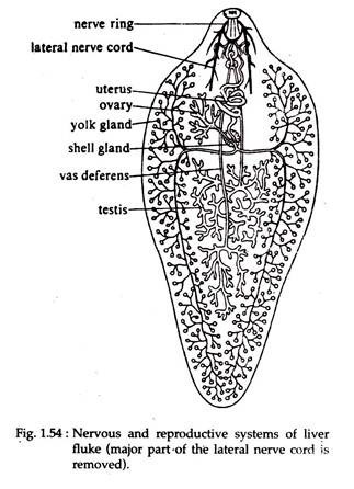
F. hepatica has a well differentiated nervous system, which also shows a bilateral arrangement. It consists of a nerve ring which surrounds the oesophagus (Fig. 1.54) and on it are present two lateral thickenings or ganglia, containing nerve cells.
From these ganglia a number of nerves are given off. Of these nerves the chief ones are a pair of lateral nerve cords running back to the posterior end and giving off numerous branches. Sense organs in Fasciola are lacking and the sluggish movement of the animal does not demand a large correlation centre. Absence of sense organ is due to parasitic mode of life.
6. Reproductive System of Fasciola Hepatica:
Fasciola reproduces sexually and their reproductive organs are constructed on the hermaphrodite plan, consisting of both male and female organs in the same individual (Fig. 1.54).
Male:
Male reproductive system consists of a pair of testes, which are in the form of a much branched tubules and occupy the middle of the posterior part of the body, one situated behind the other. From each testis there runs forward towards the anterior region a duct, the vas deferens.
The two vasa deferentia unite and form a median coiled and dilated vesicula seminalis. From it a narrow tube, the ejaculatory duct, leads to the male aperture situated at the tip of the cirrus or penis. The penis opens into the genital atrium.
Female:
The female part of the reproductive system consists of a single ovary or germarium. The ovary is in the form of a ramified tube situated in the right anterior region and above the middle line of the body, in front of the testes.
The tubules of the ovary open into a common narrow tube, the oviduct. Vitelline glands consist of numerous, minute rounded follicles, which occupy a considerable area in the lateral region of the body. On each side are two large ducts, anterior and posterior, uniting to form a single main lateral vitelline duct, one each on the right and left.
These run transversely and open into a small chamber called yolk reservoir. From it comes out a very short median vitelline duct which opens into the oviduct.
Around this junction are grouped a mass of unicellular Mehlis’ glands or ootype glands or accessory female glands which open by small ducts into the lumen of the oviduct. The lumen of the oviduct at this region is dilated and called ootype.
The uterus is a wide convoluted tube leaving the ootype. It opens in the genital atrium near the base of the cirrus. A canal termed Laurer’s canal leads from the junction of oviduct and median vitelline duct and opens externally on the dorsal surface. The role of the Laurer’s canal is to throw out excess yolk cells and possibly the eggs.
7. Life History of Fasciola Hepatica:
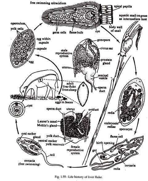
Development in F. hepatica is indirect, involving four types of free-swimming and parasitic larval stages. Fasciola is digenetic and its life cycle (Fig. 1.55) always includes at least two infective stages. Two or more hosts are infected before its life cycle is completed. The definitive or primary host is a vertebrate (sheep), while the intermediate host is a snail (Lymnaea, Planorbis etc.).
Fertilization:
Although Fasciola is hermaphrodite, self-fertilization is uncommon. During copulation, sperm exchange is mutual and cross-fertilization is the general rule. Copulation takes place in the bile duct of the host. During copulation, the cirrus or penis of one worm is inserted, via the gonopore, into the uterus of the other.
Copulation through Laurer’s canal has also been reported. Sperms are thus ejaculated. The prostate gland supplies semen for sperm survival. Sperms then travel up the uterus, through the ootype, to be stored in the seminal receptacle.
Release of Fertilized Eggs from Primary Hosts (sheep) Body:
After being released from the ovary, the eggs are fertilized either in the oviduct or within the ootype. Each egg receives a fair amount of yolk from the yolk cells and vitelline secretions. It finally becomes enclosed in a proteinaceous shell or capsule secreted by the shell glands.
The shell becomes hard when it enters the uterus. The hardening is caused by the action of quinone. One pole of the egg shell bears a small lid or operculum for the exit of the future larva. The egg thus becomes complete and remains for a little time in the uterus.
Eventually the egg leaves the fluke’s body through its gonopore and passes down the bile ducts of the sheep into the intestine, from where it is discharged to the exterior along with the faeces. The egg can survive if only it falls on damp soil.
Miracidium Larva:
Active development within the zygote begins at this stage. After three to six weeks, depending upon the temperature, the egg shell opens at the operculum and the miracidium larva emerges. The miracidium larva has a somewhat conical body, covered all over with vibratile cilia.
A distinct head lobe or apical papilla is situated at the broad end. Behind the head lobe there are two spots of pigment, the eye spots. Within the body just below the epidermis lie delicate layers of circular and longitudinal muscle fibres, the mesenchyme.
There also lies one pair of flame cells with ducts and a sac-like intestine. The rest of the interior is filled with a mass of germ balls. The head lobe bears penetration glands. The miracidium larva swims freely in water or crawls over damp surface for some time and dies unless it happens to reach a particular water snail (preferably Limnaea truncatula).
8. Infection to Secondary Host (Snail) of Fasciola Hepatica:
Sporocyst:
The miracicium larva on encountering the snail, bores into it by means of the penetration glands and reaches the internal organs, specially the pulmonary sac. It metamorphoses into the sporocyst by casting off its ciliated covering.
The sporocyst is elongated, with an internal cavity containing germ balls and lined by a layer of cells, with remnants of the eye spots and flame cells. The germ balls, eventually, undergo a process of cleavage, resulting in the formation of redia larva (first generation).
Redia Larva:
Redia is provided with mouth, pharynx and a simple intestine and there is a system of excretory vessels. It bears a circular ridge or collar near the anterior end, formed by the bulging of body wall. The fully formed body of redia is elongated and bears a pair of short, muscular projections at the posterior side.
In the interior of the body are undifferentiated germ balls and these either develop into second generation of redia if it is winter, or if it is summer, it gives rise to cercaria larva. An opening near the collar, known as the birth pore is formed in the wall of redia, through which the cercaria escapes.
Cercaria Larva:
The cercaria is provided with a long tail and with oral and ventral suckers. Alimentary canal is well developed and consists of mouth, pharynx and a bifid intestine. Paired excretory tubules with flame cells, germ balls and peripheral cyst-forming cells are also present.
They move actively by the help of their tail and forces their way out of the snails body. Then losing their tail they become encysted. The cercaria in encysted condition is called metacercaria larva.
9. Infection to New Primary Host (Sheep) of Fasciola Hepatica:
The metacercaria remains attached to blades of grass or leaves of other herbs. When the sheep feeds on this infected green leaves, metacercaria enters the gut. The young fluke emerges from the cyst and bores its way through the gut wall, passes over the viscera and penetrates into the liver and finally into the bile duct, where it grows rapidly and reaches sexual maturity.
From the life cycle of F. hepatica it can be seen that after having successfully invaded into the intermediate host (snail), Fasciola’s developmental stages undergo two rounds of asexual divisions. Once through the formation of either one or two generations of many redia and then through the formation of numerous cercariae.
This greatly increases their number and thus, they have good chances to complete their life cycle. Thus, one egg has the capability to give rise to dozens of sexual adults in the definitive host.
