Learn about the comparison of excretory system in various Arthropods.
Comparison: Arthropod # Macrobrachium:
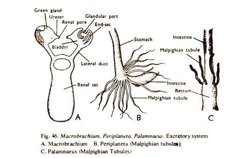
1. The excretory organs consist of a pair of cream coloured antennary glands with their ducts, a median renal sac and a transverse communicating duct. It also performs the function of osmoregulation (Fig. 46A).
2. The antennary gland is placed in the coxa of the second antenna. It consists of three parts.
(a) A small end sac.
ADVERTISEMENTS:
(b) A glandular mass. These two extract excretory products which are carried to the exterior through the bladder.
(c) The bladder occupies the innermost region and is drawn into a narrow tube to open to the exterior through the renal aperture on the inner side of the coxa. The gland is also called green gland.
3. The renal sac is a thin-walled median structure lying just above the stomach. It is connected with each antennary gland by a narrow duct anteriorly. The two ducts are again connected by a narrow transverse duct. Posteriorly, the renal sac ends blindly at the region of the gonad. The renal sac acts as a temporary reservoir for waste products.
4. No other renal organs
ADVERTISEMENTS:
5. The thick chitinous layer of the integument is a nitrogenous product secreted by the ectoderm and is cast off in each moult. Moulting is considered as a special mechanism to get rid of nitrogenous wastes.
Comparison: Arthropod # Periplaneta:
1. The excretory organs consist mainly of numerous Malpighian tubules and nephrocytes (Fig. 46B).
2. The Malpighian tubules are thread-like yellow structures of variable number present at the junction of the stomach and the intestine. Each tubule is a hollow structure opening into the lumen of the intestine and the wall is made of a single layer of glandular epithelium. Nitrogenous wastes are absorbed by the cells from the haemocoelomic fluid and the same are discharged into the lumen of the tubule which in turn conveys the wastes into the lumen of the intestine.
3. No such structure.
ADVERTISEMENTS:
4. Certain cells (nephrocytes) associated with the fat bodies and the hypodermis also serves as excretory organs.
5. The thick chitinous layer of the integument is a nitrogenous product secreted by the ectoderm and is cast off in each moult. Moulting is considered as a special mechanism to get rid of nitrogenous wastes.
Comparison: Arthropod # Palamnaeus:
1. The excretory organs consist of a few Malpighian tubules and a pair of coxal glands (Figs.46C and 47C).
2. The Malpighian tubules are delicate tubes and are two pairs in number, attached to the intestine. Each tubule is a hollow structure opening into the lumen of the intestine and the wall is made of a single layer of glandular cells. Nitrogenous wastes are absorbed by the cells from the haemocoelomic fluid and the same are discharged into the lumen of the tubule which in turn conveys the wastes into the lumen of the intestine.
3. No such structure.
4. A pair of coxal glands is found near the bases of the 3rd walking legs in the 5th segment of the prosoma. These are derived from coelomoducts and function as excretory organs. In the embryo of scorpion, the coelomoducts are found in the 3rd, 4th, 5th, 6th and 8th segments.
All of them disappear in the adult stage except those in the 5th segment which persist as coxal glands. Each coxal gland consists of an end sac, a colied tube and a bladder opening to the exterior by a duct at the base of the 3rd walking leg.
(a) A diverticulum or lymphatic organ, possibly excretory (phagocytic) in function, lies on each side of the abdomen connected with the coxal gland of the side.
5. The thick chitinous layer of the integument is a nitrogenous product secreted by the ectoderm and is cast off in each moult. Moulting is considered as a special mechanism to get rid of nitrogenous wastes.
ADVERTISEMENTS:
