The following points highlight the three main components of blood in vertebrates. The components are: 1. Red Blood Cells 2. White Blood Cells 3. Blood Platelets.
Component # 1. Red Blood Cells (RBC) in Vertebrates:
It is also called as erythrocytes.
Structure:
These are small cells of average diameter 20 pm (7-8 µm in man). All RBC have nuclei except those of mammals. Mature erythrocytes of mammals lack nuclei. In vertebrates these cells are biconvex, but due to absence of nucleus in mammals it is biconcave.
ADVERTISEMENTS:
The cell membrane is thin, through which transport of gases easily takes place. These cells function as container for haemoglobin. Haemoglobin is the major oxygen transport molecule. If it is left free in the plasma it gets excreted by the kidney. The average life span of these cells is three to four months in circulating blood.
Functions:
In vertebrates, ach RBC contains approximately 280 million haemoglobin molecules. Each haemoglobin molecule is composed of four peptide chain, called globin. These globins are bonded with an iron pigment called haeme. This haeme part attaches with oxygen in lungs and dissociates oxygen in different tissues.
The carbonic anhydrase, an enzyme present in RBC, plays important role in CO2 transport through blood. RBC lacks mitochondria; consequently it performs anaerobic respiration for its self-maintenance.
ADVERTISEMENTS:
Fate:
Erythrocytes are destroyed in liver or spleen. Its protein part is converted into amino acids. The iron molecule of haeme is used to form ferritin, and is stored in liver. These ferritins are again either used to form haemoglobin or act as a component of cytochrome molecules.
Other parts of the haeme are utilized to form different bile pigments e.g., bilirubin — a red pigment or biliverdin – a green pigment. These are utilized in the formation of bile and excreted through digestive system.
Component # 2. White Blood Cells (WBC) in Vertebrates:
These cells are also called leucocytes.
ADVERTISEMENTS:
Structure:
These are nucleated living cells and bigger in size than RBCs. They do not contain haemoglobin. These cells show amoeboid movement and their life span is shorter than other cells. These are rich in nucleoproteins and also contain lipids, glycogen, cholesterol, ascorbic acid and a variety of enzymes, especially proteolytic enzymes.
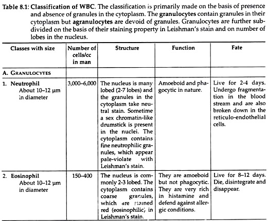
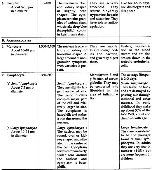
There are several varieties of leucocytes in the blood. Each variety possesses characteristic morphology and staining property in Leishman stain. Different classes of WBC are described in Table 8.1.
Functions:
1. Phagocytosis:
The neutrophils and the monocytes engulf foreign particles and bacteria and generally digest them. This process is called phagocytosis. The eosinophils and lymphocytes have got slight phagocytic function.
2. Antibody formation:
ADVERTISEMENTS:
Lymphocytes manufacture β and Ƴ fractions of serum globulin. Immune bodies are associated with Ƴ-globulin fraction.
3. Formation of fibroblasts:
Lymphocytes may be converted into fibroblasts in an area of inflammation and help in the process of repair or healing of wounds.
4. Secretion of heparin:
The basophil leucocytes are supposed to secrete heparin, which prevent intra-vascular clotting of blood.
5. Antihistaminic function:
The granulocytes, especially the eosinophil cells are very rich in histamine. They are believed to either act against or defend allergic conditions, in which histamine-like bodies are produced in excess.
6. Manufacture of trephones:
Leucocytes manufacture certain substances from plasma proteins, which exert great influence on the nutrition, growth and repair of tissues. These substances are called trephones.
Life and fate of WBCs in vertebrates:
The life of the different varieties of leucocytes differs. It is believed that granulocytes live for 1-2 days, neutrophils for 2-4 days, eosinophils for 8-12 days, basophils for 12-15 days. It is known that all the varieties of leucocytes die, disintegrate and disappear.
The neutrophils and other granulocytes undergo fragmentation in the blood stream and are also broken down by the reticulo-endothelial cells. The lymphocytes leave the body and are destroyed by passing out through the intestinal and other mucosa.
Component # 3. Blood Platelets (Thrombocytes) in Vertebrates:
Structure:
The platelets are unit membrane bound, non-nucleated, round or oval, biconvex disc like bodies. Its size varies from 2.5 µm to 5 µm in diameter. Under light microscope two components of a platelets are seen. One is the clear ground substance, called hyalomere and the other is deeply stained central chromatomere or granulomere.
The hyalomere contain microtubules and microfilaments. The microfilaments contain thrombosthenin, which can contract like actin and myosin of muscle cells. The chromatomere contains numerous cellular organelles.
The organelles are:
(i) a-granules that are 0.25 µm in diameter and always membrane bound;
(ii) Mitochondria;
(ii) Sydersomes that are iron-containing (ferritin) vesicles;
(iv) Serotonin containing granules,
(v) Glycogen granules,
(vi) Ribosomes,
(vii) System of tubules and vesicles, etc.
Functions:
1. Blood clotting:
In vertebrates, when blood is shed, the platelets disintegrate and liberate thromboplastin which activates prothrombin into thrombin.
2. Repair of capillary endothelium:
Endothelial lining of the capillaries when damaged, the circulating platelets immediately adhere in that area and quickly repair the damage. Capillary damage is a frequently occurring incident in the body. If it is not quickly repaired, the vessels will break at these spots and capillary bleeding will take place.
3. Haemostatic mechanism:
When a blood vessel ruptures blood comes out from that place. The cessation of blood flow takes place through simultaneous coagulation and agglutination of platelets in the ruptured spot.
4. Hasten clot retractions:
Speed of clot retraction is directly proportional to the number of platelets present. This retraction process is dependent upon the thrombosthenin (contractile protein of platelet) in presence of ATP and magnesium ions.
Fate:
The average life of platelets in vertebrates is about 5 to 9 days. They are destroyed in the spleen and other reticulo-endothelial cells.

