In this article we will discuss about the digestive systems of Branchiostoma and Ascidia.
Digestive System of Branchiostoma:
In Branchiostoma, the digestive and the respiratory systems are intimately associated with one another. The pharynx is a very elaborate structure which occupies more than half of the total surface area of the body. The food particles are filtered from the respiratory water current that passes in through the mouth and go out through the atriopore via the gill-slits.
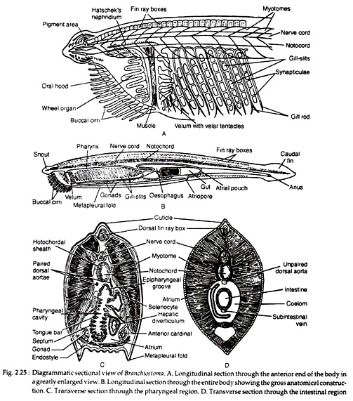
The cilia present in the pharyngeal cavity play the most important role in capturing the food. A large median aperture is situated just below the pointed tip of the anterior end. This aperture is surrounded by a frill-like membrane called oral hood (see Fig. 2.25A). The membranous lateral and ventral margins of the oral hood are fringed with buccal cirri. The buccal cirri are beset with sensory papillae.
The oral hood and the buccal cirri are supported by skeletal rods. During the inflow of water current, the buccal cirri form a sieve to prevent the entry of large particles. The oral hood encloses a cavity called vestibule. Mouth is a small rounded aperture situated at the posterior end of the vestibule.
ADVERTISEMENTS:
It is surrounded by a membrane called velum. Velum is produced into 12 slender velar tentacles (or languets). It acts as a sphincter to open and close the mouth (see Fig. 2.25A, B). The lining of the vestibule produces a complicated ciliated tract called wheel organ or rotatory organ or ciliated organ of Muller.
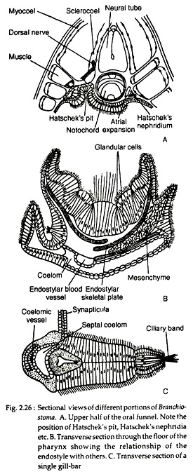
The wheel organ produces a vortex of water to sweep the food matters into the mouth. There is a ciliated glandular groove running along the roof of the vestibule named the groove of Hatschek which ends into a small depression called the pit of Hatschek (Fig. 2.26A). The left embryonic coelomic cavity of the head region becomes reduced to form the pit of Hatschek in the adult.
The mouth opens into a very large laterally compressed tube called pharynx. The pharynx in Branchiostoma plays a dual role — collection of food particles and respiration. The lateral wall of the pharynx is perforated by obliquely arranged vertical apertures — the gill-slits (see Fig. 2.25B).
ADVERTISEMENTS:
The number of gill-slits is about 200, but the gill-slits increase far beyond the number of body segments as the animal grows older. The gill-slits open into a special cavity, the atrium or peribranchial cavity, which surrounds the pharynx on all sides except the dorsal. The atrium is closed anteriorly but opens to the exterior at the posterior end through the atriopore (Fig. 2.25B-D).
The gill-slits are separated from one another by portion of walls of the body and the pharynx. These portions are called the gill- bars or branchial lamellae, i.e., the gillbars are actually the vertical portions of the main body wall and the pharyngeal wall. Such a portion of the gill-bar which encloses the coelom is called primary gill-bar.
With the advancement of age, each primary gill-slit is divided into two by the downward growth of the tongue bar. These resultant gill-bars are named as the secondary gill-bars which lack coelom. The gill-bars are provided with cilia. The gill-bars contain supporting gill-rods. The gill-rods are of two types — the primary gill-rods and the secondary gill-rods (Fig. 2.25A).
ADVERTISEMENTS:
The terminal end of the primary gill-rod is forked and those of the secondary gill-rods are simple, i.e., unforked. Depending on the presence of particular type of gill-rods, the gill-slits are also designated as the primary or secondary ones. The primary branchial lamellae are connected by transverse skeletal rods called the synapticulae (Fig. 2.26C).
The internal wall of the pharynx is ciliated. Several ciliated tracts are present inside the pharynx. An endostyle is present on the floor of the pharynx. The endostyle consists of a few tracts of ciliated cells alternating with mucus-secreting glandular bands (Fig. 2.26B).
According to Barrington (1968), the endostyle becomes transformed into thyroid gland in case of vertebrates. He has observed the concentration of radioactive iodine in the glandular columns of the endostyle. The extract contained iodine and, on administration of it, acceleration of the metamorphosis of tadpole larva occurred.
There is another median groove called epipharyngeal or hyper pharyngeal groove on the dorsal side of the pharyngeal cavity. The epipharyngeal groove joins with the anterior end of the endostyle by peripharyngeal ciliated tracts.
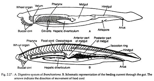
Behind the pharynx, the alimentary canal narrows at its posterior side into an esophagus which opens into the midgut. The pharynx and oesophagus constitute the foregut. At the junction of the oesophagus and the midgut there is a large out pouching called hepatic diverticulum or midgut diverticulum (Fig. 2.27A).
The diverticulum extends forward on the right-hand side of the pharynx for about one-third of the length of the pharynx. It contains zymogen cells and produces digestive enzymes. The inner walls of the diverticulum, specially the dorsal and ventral walls are beset with large cilia.
The posterior end of the midgut is specially ciliated and called ileocolon ring. Behind the ileocolon ring, the, intestine proceeds backwards as a straight hindgut which opens through the anus.
Mechanism of feeding and digestion in Branchiostoma:
ADVERTISEMENTS:
Branchiostoma is a microphagus animal which obtains the food by filtering the stream of water that enters the pharyngeal cavity. The food or ‘sea-soup’ consists of protozoans, algae, diatoms and other organic particles suspended in sea-water. The wheel organ produces a vortex of water current which is drawn in towards the mouth.
The buccal cirri become curved to form a sieve to prevent the entry of large particles. The sensory papillae in the buccal cirri and velar tentacles act as chemoreceptors and taste the nature of the food particles and also estimate the size of food particles. If food particles are large in size or liable to cause toxicity, these are expelled by the forceful expulsion of the water from the pharyngeal cavity.
The ingress of water into the pharyngeal cavity through the mouth is controlled by the velum. The pharynx plays the most important role in food collection. The major portion of the water passes out into the atrium through the gill-slits. The cilia present on the gill-bars beat to drive the water out into the atrium and, thus, facilitate the inflow of fresh water current through the mouth.
The food particles, due to their own weight, begin to fall on the floor of the pharyngeal cavity and are entangled by the sticky secretion of the mucus-secreting cells of the endostyle. The cilia in the endostyle and gill-bars beat to produce an upward current to push the mucus entangled food particles towards the epipharyngeal groove.
The cilia of the endostyle also beat to drive the food along the peripharyngeal ciliated tracts to the epipharyngeal groove. The food is then pushed backwards by the backward beating of the cilia of the epipharyngeal groove (Fig. 2.27B). The secretion of the glandular cells of the endostyle transforms the boluses of mucus-entangled food particles into a cord-like structure known as food cord.
The food cord from the pharynx passes through the oesophagus into the hepatic diverticulum and midgut where this food cord is subjected to the action of digestive enzymes secreted by the hepatic diverticulum. The food cord from the hepatic diverticulum is pushed backwards by the cilia present in its cavity. The mucus-entangled food cord is rotated by the ciliary action in the ileocolon ring.
Digestion in Branchiostoma is both intracellular as well as extracellular. The intracellular digestion takes place inside the hepatic diverticulum while the extracellular digestion occurs inside the midgut. The secretory cells of the hepatic diverticulum contain zymogen granules and they show phagocytosis, i.e., the cells are able to engulf the food particles from the food cord and digest the food as seen in Amoeba and Hydra.
The phenomenon of phagocytosis is attested by the fact that carmine particles, after injection into the diverticulum, are taken inside the cells. The digestive enzymes in Branchiostoma are amylase, lipase and protease. The digested food is absorbed in the hindgut and the undigested particles are expelled through the anus.
The controlling mechanism of the ciliary mode of feeding in Branchiostoma is not clearly known. The afferent and efferent nerve fibers in the atrium presumably play the important role in feeding. The rate of water current is largely controlled by the intensity of beating of cilia and also the degree of contraction or dilatation of the inhalant and exhalant apertures.
The different receptors present on the velum and the atrium taste the nature of water current. If the water current contains any toxic substance, the atriopore closes and the water is regurgitated by sudden contraction of the pterygial muscles which form the floor of the atrium.
Digestive System in Ascidia:
In Ascidia, mouth is situated at the centre of the velum placed in the posterior end of the oral funnel. The velum which partially separates the pharynx from the mouth is provided with velar tentacles. The opening and closure of the mouth are regulated by muscle bands. The mouth leads into a very short stomodaeum which opens into a greatly enlarged sac-like pharynx or branchial chamber.
The pharynx in Ascidia performs two functions — one is food collection and the other is respiration. The pharynx extends nearly up to the base of the body and remains attached with the mantle along the ventral side.
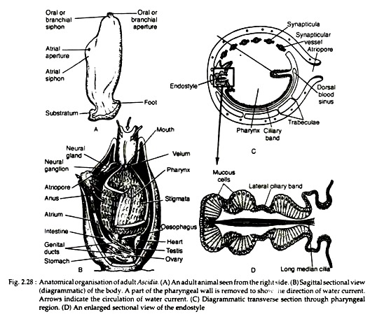
Except this region the whole of the pharynx is enclosed by a special cavity called the atrium (Fig. 2.28B). Atrium opens to the exterior through an atriopore. The pharyngeal wall and the body wall are connected by trabeculae (Fig. 2.28C).
The wall of the pharynx is pierced by numerous vertically disposed apertures called stigmata. The stigmata develop by the subdivision of a few (presumably three) gill-slits. Each gill-slit is divided by the developing tongue-bars which become connected by transverse connecting bars called synapticulate. By this way numerous stigmata are produced out of a few gill-slits. The stigmata bears series of papillae containing muscles and cilia.
An endostyle is present in the ventral side of the pharynx. The endostyle has two rows of mucus cells (Fig. 2.28D) on each side which are separated by row of ciliated cells. A group of median cells with very long cilia are present at the middle of the endostyle (Fig. 2.28D).
Experimental studies with the isotopes of iodine show that certain cells on the glandular tracts of endostyle incorporate iodine. The opposite side of the endostyle is occupied by the dorsal lamina which is produced into numerous hanging curved bodies called languets (Fig. 2.28C). The endostyle and the dorsal lamina are connected by the peripharyngeal ciliary bands.
The pharynx is connected with an expanded stomach by a short and narrow oesophagus (Fig. 2.28B). The stomach has a fusiform shape and its folded inner wall contains digestive glands. These glands secrete digestive enzymes like protease, lipase, amylase and invertase. The gastric secretion has a strong carbohydrate-splitting enzyme and weaker lipolytic and proteolytic enzymes.
There is no liver. A pyloric gland composed of numerous small sacs or ampullae remains in contact with the intestine. Ductules arising from these ampullae unite to form a duct. This duct opens into the posterior end of the stomach. The physiological role of pyloric gland is not fully ascertained. Both digestive and excretory functions have been suggested by many workers.
From the stomach, a simple intestine ascends up and opens into the atrial funnel by the anus. The wall of the alimentary canal is non-ciliated. The intestine is the absorptive portion of the alimentary canal. The wall of the intestine is thickened internally into a pad called typhlosole which increases the absorptive surface of the intestine.
Food collection:
The beatings of cilia in the stigmata set up a water current which enters into the pharynx and goes out through the atriopore via the stigmata. The food particles suspended in the water current are entangled by the mucus secreted by the glandular tracts of endostyle. The mucus-entangled food particles move upwards and are then pushed back into the oesophagus by the ciliary action of the dorsal lamina.
From the oesophagus, the food particles pass on into the stomach where they are subjected to the action of the digestive ferments. The digested food is absorbed in the intestine and the undigested products are expelled out through the anus and atriopore.
4. Stomach in Mammals:
The stomach in mammals is transversely arranged and in most forms they are typical. In platypus the two limbs — namely cardiac and pyloric — are almost fused along the lesser curvature and appear as a wide sac. Since the lining of epithelium is devoid of glands the stomach in monotremes is not considered as a true stomach.
The cardiac and pyloric regions in some monkeys and rodents are marked by a constriction in between them. Such stomach is known as hour-glass stomach. The pyloric part of the stomach of kangaroo is provided with many peculiar and sacculated folds in its wall. In the blood sucking bat, Desmodus, the pyloric part has become elongated to form a caecum-like structure for storage of sucked blood.
Ruminant stomach:
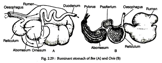
Stomach of ruminating mammals is very complex. The stomach of Ruminantia is constituted of four separate chambers. These chambers are known as rumen (paunch), reticulum (honeycomb) omasum (psalterium or manyplies) and abomasum (rennet or reed) (Fig. 2.29). Of these, the first three are considered by many as modified parts of the oesophagus but embryological studies show that they are really parts of stomach.
These parts are lined by stratified epithelium and folded into muscular ridges. The rumen is large and saclike and serves mainly as storage organ. Muscular ridges are low in rumen, form a network in the reticulum and are over-lapping leaves in the omasum. The cattle feed fairly rapidly and fills the rumen with grain, grass and other herbage.
The food within the rumen is churned by muscular contraction and undergoes certain bacterial fermentation during its stay in the rumen. Organic acid, from acetic acid upwards, are produced, absorbed into the circulation and metabolised. Bacteria and protozoans, which digested cellulose and are themselves later digested.
Food from the rumen passes by degrees into the reticulum or directly to the oesophagus and, hence, to the mouth by a process of retro peristalsis. Such food regurgitated into the mouth is called cud or bolus. When the cud is well-masticated and thoroughly mixed with the secretion of the salivary glands, it again passes into the rumen.
A new bolus is again formed and chewed and sent back to the rumen. The process is repeated several times and when all the food is well-masticated it passes to the reticulum and then to the omasum. Here water is pressed out and absorbed and remainder proceeds to the abomasum. The abomasum is provided with gastric glands and considered as ‘true stomach’.
Camel’s stomach:
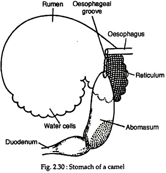
The true ruminant stomachs occur under the suborder Ruminantia. The camel’s stomach is different in anatomy and histology from the stomach of ruminants, but their rumination and fermentation are the same like the true ruminants.
In camels the stomach is three-chambered, the omasum being absent. The rumen and reticulum parts of the stomach of camels are provided with pouch-like water cells (Fig. 2.30). Their openings are guarded by sphincter muscles. At one time it was believed that the camels store water in their water cells and make prolonged journey without drinking water.
But the view is no longer tenable. The pouches can by no means held as much water as would be required by the animal on a prolonged journey through the desert where no water is available. Later studies have shown that the camels get their water requirement during journey by the breaking down of the glycogen of the muscles and fat of the hump.
Physiologists have shown that for each 100 gm. of fat oxidised in the body 110 gm. of water is formed. They also have shown that the water cells do contain some metabolic water. The water is pure in nature and helps in moistening the food. Abomasum is provided with gastric cells and acts as true stomach.
Stomach of some other mammals:
The stomach of whales and hippopotamus is also divided into several chambers.
The glands that are found in the stomach have been named according to their location.
The glands located on the cardiac part of the stomach are called cardiac glands. Those located on the fundus and pyloric parts are called fundic and pyloric glands, respectively. The fundic glands are of great importance in digestion. There are three types of fundic glands. They are mucous neck cells, chief or zymogen cells and parietal or acid-secreting cells.
The secretion of the stomach is known as gastric juice. It contains mucus, hydrochloric acid, pepsin, renin and gastric lipase. The concentration of gastric hydrochloric acid in human gastric juice varies from 0.05 to 0.3 per cent. In dog this concentration may be as high as 0.6 per cent.
