In this article we will discuss about:- 1. Integument in Mammals 2. Horns in Mammals 3. Antlers.
Integument in Mammals:
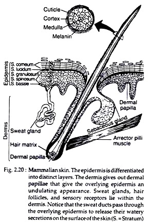
Epidermis is divided into five layers; from outside these are as follows (Fig. 2.20):
(i) Stratum corneum:
ADVERTISEMENTS:
This is the outermost keratin layer. Cell boundary is indistinguishable and nucleus is absent. This layer is thick in the palm and sole and thin in the lips. Hair developed from this layer.
(ii) Stratum lucidum:
This is the second layer from the outside. It is a thin layer. Cells are devoid of nucleus and cell membrane is inconspicuous. The cells of this layer possess eleidin which gives rise to keratin.
(iii) Stratum granulosum:
ADVERTISEMENTS:
Third layer from the outside and is composed of three to five cell layers. The cells contain keratohyaline granules, responsible for the name of the layer.
(iv) Stratum spinosum:
This is fourth layer from the outside. It is much thicker than the others. The cells of this layer bear several spiny structures emerging out from the membrane. So these are called prickle cells. These spines attach with the spines of the adjacent cells.
(v) Stratum basale:
ADVERTISEMENTS:
This is the fifth and last layer of the epidermis. The cells of this layer are columnar and continuously dividing mitotically to replace upper layers. Cytoplasmic extensions from the basal cells reach the cells of dermis.
(vi) Stratum Malpighi:
This layer is not the part of epidermis. But present in-between epidermis and dermis. This layer possesses branched cells called melanocytes. Dermis or Cutis vera is composed of collagenous and elastic fibres. Upper layer of dermis is well-organised papillary layer, which maintains connection with the melanocytes and prickle cells.
Inner layer of dermis is loose connective tissue with fat deposition. Fibroblast cells of the dermis produce fibrous tissue and reticulo-endothelial system. Some of the cells possess melanin pigment and are called melanophore. Dermis contains sweat, ceruminus, and sebaceous glands.
Horns in Mammals:
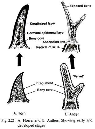
Horns appear as outgrowths of the skull beneath the integument, which forms a keratinized sheath. Horns occur in bovids of both sexes and are usually retained year round. The horns are found only in certain members of the order Artiodactyla and in Rhinoceros of the order Perissodactyla. According to structure and mode of formation, three types of horns are found in mammals [Fig. 2.21A].
1. Keratin fibre horn:
In case of Rhinoceros a hard conical structure is present on the front of the nasal region of the skull. It is composed of a cluster of bony fibres. The fibres are fused together by a mass of hard and keratinized cells growing from the epidermis. Each fibre resembles a very thick hair and emerges from a dermal papilla. The fibres are, however, not true hair as their bases lack follicles.
2. Hollow horn:
ADVERTISEMENTS:
In artiodactyls a pair of horns are present, one on each of the frontal bone (Fig. 2.21). The horn consists of a bony projection from the frontal bone of the skull. A cornified layer of epidermis covers the projection. A cavity enters into the bony projection. The horny layer is never shed in the lifetime.
3. Prong horn:
It is a conical projection on the frontal bone. A horny epidermal sheath covers the projection. The sheath usually bears prong. This unique type of horn is found in antelope, Antelocopra americana of Western America (Fig. 2.22).
Antlers in Mammals:
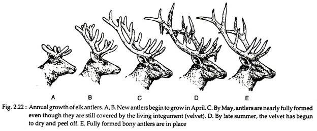
These are mesodermal derivatives. In natural condition these are hard bony structures develop on the frontal bone of the skull. Antlers appear as outgrowths of the skull beneath the overlying integument, which is referred to as “velvet” because of its appearance. This overlying velvet dries and falls away, leaving the bony antlers.
Antlers are restricted to members of the deer family and, except for caribou (reindeer), they are present only in males. Antlers are sheded and replaced annually [Fig. 2.21B and 2.22]. A mature antler has many branches, e.g. males of some deer, both male and female of giraffe and caribou possess antlers. In giraffe the skin covering of the antlers never shaded; it remains throughout life period.
Hoof, claw and nails in mammals:
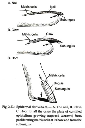
In ungulate mammals the end of the digits remains covered by thick unguis, called hoof. Sub-unguis is present below the unguis (Fig. 2.23).
In case of claws and nails, sub-unguis plate is rudimentary and attached with the pad of the digit. The unguis is sharp curved needle- like. The members of the feline group can withdraw their claws within the paws. In nail the unguis is broad and flat; sub-unguis is rudimentary and remains at the base of the nail.
Hair in mammals:
Hair are slender keratinous filaments. Root is the base of a hair, the remaining length constitute shaft. The outer surface of the shaft forms a scaly cuticle. Beneath this, is the hair cortex, and at its core is the hair medulla (Fig. 2.20).
The hair shaft is produced in the epidermal hair follicle and projects above the surface of the skin. The surface of the epidermis dips down into the dermis to form the hair follicle. At its expanded base, the follicle receives a small tuft of the dermis, the hair papilla. The arrector pilli muscle, a thin band of smooth muscle anchored in the dermis, is attached to the follicle and makes the hair stand erect in response to cold, fear or anger.
Thick covering of hair is fur or pelage, generally composed of guard hair and under fur. The larger, coarse hair, called guard hair, are the most apparent on the outer surface of the fur. The under fur is situated beneath the guard hair and is usually much finer and shorter.


