In this article we will discuss about the Endoplasmic Reticulum:- 1. Meaning of Endoplasmic Reticulum 2. Morphology of Endoplasmic Reticulum 3. Chemical Composition 4. Biogenesis 5. General Functions.
Contents:
- Meaning of Endoplasmic Reticulum
- Morphology of Endoplasmic Reticulum
- Chemical Composition of Endoplasmic Reticulum
- Biogenesis of Endoplasmic Reticulum
- General Functions of Endoplasmic Reticulum
1. Meaning of Endoplasmic Reticulum:
Endoplasmic reticulum (ER) composes a system of membranes that enclose a space. The fluid content of cytoplasm is accordingly divided by the ER into two compartments the space enclosed within the membranes, which is termed as the luminal or cisternal space, and the region outside of the membranes which is the cytosolic space.
ADVERTISEMENTS:
These spaces or cavities often remain concentrated in the endoplasmic portion of the cytoplasm and therefore, is known as endoplasmic reticulum — ‘a net in the cytoplasm’ (Eighteenth-century European ladies carried purses of netting called reticules). The name ‘endoplasmic reticulum’ was first coined by Porter in 1953, who in 1945 had observed it in liver cells under electron microscope.
The occurrence of ER varies from cell to cell. In hepatocytes, both SER (smooth ER) and RER (rough ER) are present. The erythrocytes, egg and embryonic cells lack ER. The adipose tissues, brown fat cells, adrenocortical cells and endocrine cells of testes and ovaries contain only SER.
On the other hand, cells of organs involved actively in protein synthesis (e.g., acinar cells of pancreas, goblet cells, cells of some endocrine glands) are found to contain RER that are highly developed. The size of ER varies considerably in different cell types and is related to their functions.
2. Morphology of Endoplasmic Reticulum:
ADVERTISEMENTS:
Morphologically ER is divided into two broad categories the rough endoplasmic reticulum (RER) and the smooth endoplasmic reticulum (SER). Attached ribosomes are present in former but absent in the latter.
Both types of ER may occur in the following forms:
i. Cisternae:
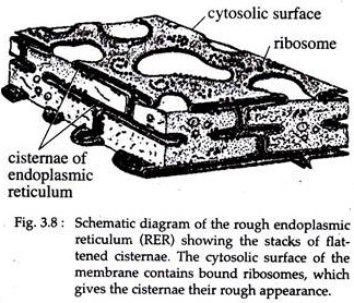
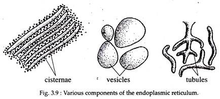
The cisternae are long, flattened, sac like, un-branched tubules, arranged parallely in bundles or stacks. Each sac is separated by a cytosolic space. RER usually exists as cisternae.
ADVERTISEMENTS:
Although the cisternal space of the RER appears in electron micrograph to be divided into separate compartments, it is thought that all of the RER cisternae communicate with one another and that the cisternal space is continuous among them (Fig. 3.8).
ii. Tubules:
The membranous elements of SER are typically tubular. It forms an interconnecting system of pipelines curving through the cytoplasm in which they occur. Tubular form of ER is dynamic in nature, i.e., it is associated with membrane movements, fission and fusion between membranes of cytocavity network (Fig. 3.9).
iii. Vesicles:
Following homogenization of cells, the SER fragments into smooth- surfaced vesicles, whereas the RER fragments into rough-surfaced vesicles. These two types of vesicles have different densities, indicating different nature of their contents. Vesicles may often remain present in the cytoplasm of most cells (Fig. 3.9).
Rough Endoplasmic Reticulum (RER):
RER is bounded by rough walls because ribosomes remain attached with its outer surface. The ribosomes are present as polysomes held together by mRNA and are often arranged in typical ‘rosettes’ or ‘spirals’. The ribosomes appear to be attached to the membrane by large 60 subunit.
ADVERTISEMENTS:
The cavity of RER is sometimes very narrow, with two membranes closely apposed; but more frequently there is a true space between the membranes that may be filled with a material of varying opacity. The cells that are actively engaged in protein synthesis, such as plasma cells and goblet cells possess a dense macromolecular material inside the cisternae.
Isolated RER contains two trans-membrane glycoproteins, such as ribophorin I and ribophorin II of molecular weighs 65,000 and 64,000 Daltons, respectively. These proteins tend to interact with each other arid form intramembranous network, which control the ribosome binding sites in the plane of the ER membrane. In general, RER is highly developed in cells of pancreas, liver, goblet cells, plasma cells etc.
Smooth Endoplasmic Reticulum (SER):
The smooth ER though forms a continuous system with the RER, has a different morphology. It’s membrane is smooth and lack ribosomes. In general, SER are found in cells that are active in glycogen and lipid metabolism, such as, adipose cells, interstitial cells, glycogen storing cells of liver, spermatocytes and leucocytes etc.
In liver cells, SER consists of a tubular network that pervades large regions of the cytoplasmic matrix, rich in glycogen and looks as dense particles called glycosomes.
Under the electron microscope, these glycosomes appear to be spheroidal in shape, with a diameter of 50-200 nm and show an internal structure made of smaller particles of about 20-30 nm. Many glycosomes are also found to be attached to SER in the conduction fibre of the heart.
3. Chemical Composition of Endoplasmic Reticulum:
By differential centrifugation, most of the components of the endomembrane system can be isolated in the so called microsomal fraction called microsomes or closed vesicles of either a rough or smooth form, that are not found in the intact cells.
In rat liver, the membranes of microsomes contain 60 to 70 per cent protein and 30 to 40 per cent phospholipid by weight. Thus, ER membrane contains more proteins, both in quantity and quality than the plasma membrane.
The membranes of ER are found to contain many enzymes, triglycerides, phospholipids and cholesterol. Most important ER enzymes include stearates, NADH-cytochrome C reductase, NADH-diaphoresis, glucose-6-phosphatase, glycosyl transferase and Mg++ activated ATPase. The various enzymes have different topologies with respect to the luminal or cytoplasmic surfaces of the ER.
4. Biogenesis of Endoplasmic Reticulum:
According to current concepts, membrane biogenesis is a multi-step process that involves the synthesis of a basic membrane of lipids and intrinsic proteins. Then the addition of the other components, such as enzymes, sugars or lipids occur in a sequential manner. The insertion of protein into ER membrane is independent of that of the lipids.
Some proteins of the ER are formed by the ribosomes in the cytosol which then become inserted into the membrane. In ER, the phospholipids are distributed more rapidly between two monolayers than that into the plasma membrane.
The biogenesis of ER is not definitely known. As membranes of ER resemble the nuclear membrane and plasma membrane, and also at the telophase stage the ER membranes are found to form the nuclear envelope, it is assumed that ER develops by evagination from the nuclear envelope. However, the synthesis of membranes follows the direction RER → SER.
5. General Functions of Endoplasmic Reticulum:
Within the cell the ER act as a circulatory system for intracellular circulation of various substances.
The route of circulation is as follows:
RER → SER → Golgi complex → lysosome or transport vesicles or secretory granules. In this route, various particles, molecules and ions may be carried into and- out of the cells. Export of RNA from nucleus to cytoplasm may also occur by this circulation.
The membranes of ER are found to conduct infra-cellular impulse, e.g., the sarcoplasmic reticulum of muscle transmits impulses from the surface membrane into the deep region of the muscle fibre. Exchange of molecules by the process of diffusion and active transport may take place across the ER membrane.
As in plasma membrane, the ER membranes possess carriers and permeases that are involved in active transport. By dividing the fluid content of the cell into compartments, the ER provides skeletal framework to the cell and thus gives mechanical support for the colloidal matrix of the cytoplasm.
A. Special Functions of SER:
i. Synthesis of Lipids:
Phospholipid biosynthesis is largely confined to the membranes of SER. Studies with radio-active precursors indicate that the newly synthesized phospholipids are rapidly transferred to other cellular membranes by the help of specific phospholipid exchange proteins.
ii. Glycogenolysis and Blood Glucose Homeostasis:
SER is found to be related to glycogenolysis or breakdown of glycogen. In prenatal liver cells, the glycogen depletion is accompanied by an increase in SER. On the other hand, glucose-6-phosphatase present in the SER membrane removes the phosphate group, generating glucose molecules from glucose-6-phosphate that ultimately move into the blood for the maintenance of the functions of red blood cells and nerve tissues.
iii. Steroid Metabolism:
Many enzymes present in membrane of SER have a key role in the synthesis of cholesterol, the precursor of steroid hormones and bile acids.
iv. Detoxification:
SER helps in detoxification of a wide variety of zenobiotics (toxic materials of both endogenous and exogenous origin) in the liver such as, barbiturates, ethanol, aspirin and petroleum products.
Detoxification is carried out by a system of oxygen transferring enzymes (oxygenases), which include the cytochrome P450s. These enzymes lack substrate specificity and are able to oxidize innumerable hydrophobic compounds into more hydrophilic, more readily excretable derivatives.
However, these enzymes may also convert the harmless compound benzo [a] pyrene into a potent carcinogen. The regulated release of Ca++ from SER membranes triggers specific cellular responses including, the contraction of skeletal muscle cells.
B. Special Functions of RER:
i. Synthesis of Proteins on Membrane Bound Ribosomes:
Proteins that are secreted from the cells, or act as integral membrane proteins or proteins of certain organelles like Golgi complex, lysosomes, endosomes are assembled on ribosomes attached to the outer surface of RER membranes.
These proteins contain a signal sequence of 6 to 20 non-polar amino acid residues. It targets the nascent polypeptides for the ER membrane and leads to the compartmentalization of the polypeptide within the ER lumen.
A signal recognition particle (SRP), recognizes the signal sequence and acts as a ‘tag’ that allows the SRP-ribosome-nascent polypeptide to bind to an SRP receptor located on the cytosolic surface of the ER membrane.
Thus, these proteins, instead of passing into the cytoplasm, appear to pass into the cisternae of the RER, and are protected from action of cytoplasmic protease enzyme. As one nascent polypeptide enters the RER cisternae, it acts upon by a variety of enzymes located either within the membrane or in the lumen of the RER. These enzymes transform the nascent proteins into their proper functional state.
Integral membrane proteins, such as glycophorin and a fraction of the erythrocyte plasma membrane are synthesized on membrane bound ribosomes and trans-located through the ER membrane as the polypeptides elongate.
Integral proteins contain one or more stretches of hydrophobic amino acids that serve as stop-transfer sequences, which block further movement of these proteins into the cisternal chamber, but trigger the opening of the channel laterally and cause the insertion of the hydrophobic helix into the lipid bilayer.
ii. Synthesis of Membrane Lipids:
The enzymes involved in the synthesis of phospholipids are themselves integral proteins of the RER membrane with their active sites facing the cytosol.
iii. Glycosylation:
Nearly all the proteins produced on the membrane bound ribosomes are glycoproteins. Glycosylation or addition of carbohydrate chains to the proteins occur by either N-linkages or O- linkages. The formation of the carbohydrate sequences of glycoproteins occur without the use of a template.
The addition of sugars is catalyzed by a group of membrane-bound enzymes called glycosyl-transferase. If a glycoprotein is to contain a terminal glucose, fucose or sialic acid, then these sugars are added into the Golgi apparatus where the required transferase enzymes are present.
iv. Maintenance of Membrane Asymmetry:
The asymmetry of membrane is initially established in the RER. Components situated at the cisternal surface of the ER membrane can be identified on the luminal surface of transport vesicles and the luminal surface of Golgi cisternae, but the external (exoplasmic) surface of the plasma membrane.
For example, the carbohydrate chains, which are first added in the ER, are always present on the exoplasmic side of the plasma membrane following the fusion of vesicles with the plasma membrane.
v. Vesicular Transport:
The cisternae of the RER are typically large with flattened cavities that can act as transport channels for membrane and luminal proteins to be moved from their site of synthesis to the apical tips of the RER.
The apical edges of the RER cisternae are typically smooth surfaced i.e., devoid of ribosomes and are referred to as transitional elements, which serve as sites of formation of the first set of transport vesicles in the biosynthetic pathway leading towards Golgi complex.

