In this article we will discuss about the Golgi Complex:- 1. Meaning of Golgi Complex 2. Morphology of Golgi Complex 3. Chemical Composition of Golgi Complex 4. Origin of Golgi Complex 5. Functions of Golgi Complex.
Contents:
- Meaning of Golgi Complex
- Morphology of Golgi Complex
- Chemical Composition of Golgi Complex
- Origin of Golgi Complex
- Functions of Golgi Complex
1. Meaning of Golgi Complex:
The Golgi apparatus, like the endoplasmic reticulum, is a canalicular system with sacs that performs some important cellular functions like biosynthesis of polysaccharides and packaging of cellular products.
ADVERTISEMENTS:
In 1898, an Italian biologist, Camillo Golgi discovered a dark yellow network located near the nucleus of nerve cells. This network, which was later identified in other cell types, was named the Golgi complex or Golgi body. Since originally these were known to be networks, they were also called “dictyosomes” (Gr., dictyes = net).
The Golgi complex occur in all cells except the prokaryotic cells and some eukaryotic cells like mature sieve tubes of plants, mature sperm and red blood cells of animals. In animal cells, there usually occurs a single Golgi apparatus however, its number may vary.
Thus, Par-amoeba species has two Golgi apparatuses, and nerve cells, liver cells and chordate oocytes have multiple Golgi apparatuses. In animal cells, the Golgi apparatus is a localized organelle. Usually it remains polar and occurs in-between the nucleus and the periphery (e.g., thyroid cells, goblet cells).
In nerve cells it occupies a circum-nuclear position. However, in the cells of higher plants, the Golgi bodies or dictyosomes are usually found scattered throughout the cytoplasm.
ADVERTISEMENTS:
ADVERTISEMENTS:
2. Morphology of Golgi Complex:
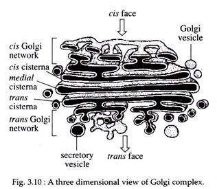
The Golgi apparatus is highly pleomorphic. In some cells it is compact and limited, in others it is reticular. Typically, however, it consists of flattened disk like cisternae with dilated rims and associated vesicles and tubules (Fig. 3.10).
The detailed structures of the basic components of the Golgi apparatus are as follows:
Cisternae:
ADVERTISEMENTS:
The cisternae are flattened, plate or saucer-like closed compartments. These are arranged in an orderly stack, much like a stack of pancakes. Typically, a Golgi stack contains fewer than eight cisternae. Depending on the cell type, an individual cell may contain from a few to several thousand stacks per cell.
The diameter of cisternae varies from 0.5 to 1.0 nm and in each stack, cisternae are separated by a space of 20 to 30 nm, which may contain rod-like elements or fibres. Each cisterna is bounded by a smooth unit membrane and is curved in a manner resembling a shallow cup.
The cisterna closest to the endoplasmic reticulum (ER) is usually convex and is said to be at the cis face or proximal face or forming face, while the cisterna at the opposite end of the stack is of concave shape and said to be at the trans or distal or maturing face. This polarization is called cis-trans axis of the Golgi apparatus.
Functionally the Golgi complex is also divided into four distinct compartments, the cis, medial, trans cisternal, and the trans Golgi network (TGN). Newly synthesized membrane, secretory and lysosomal proteins leave the ER and enter the Golgi through its cis face and then pass across the stack to the trans face (Fig. 3.10).
Tubules:
The cis or forming face is characterized by the presence of small transition vesicles or tubules that converge upon the Golgi cisternae, forming a kind of fenestrated plate.
Vesicles:
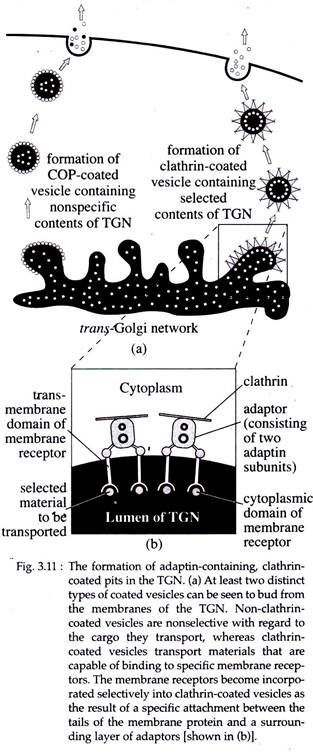
Vesicles are those that bud from the ER or from the cis and medial cisternae of the Golgi complex and are seen in the electron microscope to be covered by a indistinct fuzzy coat that contains a GTP- binding protgin, called ARF (adenosylation ribose factor).
ADVERTISEMENTS:
In addition to ARF, the coat vesicles contain a complex of seven distinct, but related coat proteins (COPs). Vesicles coated with COPs are referred to as non-clathrin-coated vesicles.
Another type of vesicles called clathrin-coated vesicles contain:
(a) An outer honeycomb like lattice composed of the protein clathrin and
(b) An inner shell, composed of protein complexes called adaptors.
Two distinct types of clathrin-coated vesicles can be distinguished, one type that buds from the TGN of the Golgi stack and contain γ-adaptin (Fig. 3.11) and the other buds from the plasma membrane, contain a-adaptin (Fig. 3.11).
The GERL Region:
The TGN or trans-Golgi network is also referred to as GERL (Golgi + smooth ER + lysosome). It is found to be involved in the origin of primary lysosome and melanin granules. It helps in processing, condensing and packaging of secretory material in endocrine and exocrine cells and in lipid metabolism. GERL is also a region of sorting of cellular secretory proteins.
Zones of Exclusion:
The Golgi complex is surrounded by a differentiated region of cytoplasm where ribosomes, glycogen and organelles, such as mitochondria are scarce or absent. This is called zone of exclusion. Coated vesicles are restricted to this region.
3. Chemical Composition of Golgi Complex:
Studies carried out on isolated Golgi complex from different plant and animal cells show marked differences in its chemical composition. The Golgi, isolated from rat liver consists of about 60% protein and 40% lipid. In animal cells, Golgi complex contains phospholipids in the form of phosphatidylcholins, whereas that of plant cells contains phosphatidic acid and phosphatidyl- glycerol.
The Golgi apparatus also contains a variety of enzymes, such as glycosyl transferases (e.g., sialyl transferases), oxireductases (e.g., NADH-cytochrome C-reductase), phosphatases (G-6-phophatase), phospholipase (phospholipase A), kinases (casein phosphokinases) etc.
Both animal and plant cells have some carbohydrate components in common, such as glucosamine, galactose, glucose, mannose and fucose, but plants also have some other special sugars.
Cytochemical Properties:
Different parts of Golgi apparatus show different histochemical staining properties. The cis face of Golgi apparatus stains positively with osmium tetroxide (OsO4), while trans face stains selectively with phosphotungstic acid. Acid phosphatase enzyme is cyto-chemically marked in the GERL region. Transferase enzymes are found to be located in the membrane of Golgi, not in the lumen of cisternae.
4. Origin of Golgi Complex:
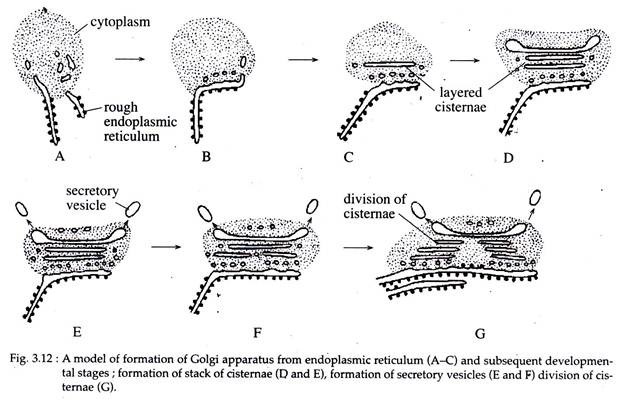
Various cytological and biochemical evidences have established that the membranes of the Golgi apparatus have originated from the membranes of the SER which in turn have originated from the RER. The transitional vesicles originated as blebs from the ER, migrate to cis-Golgi where, by coalescence, they form new cisternae.
At the distal face, the cisternae ‘give their all’ to vesicle formation and disappear. Thus, Golgi cisternae are constantly and rapidly renewed. Individual cisterna, however, may arise from the pre-existing stacks by division or fragmentation (Fig. 3.12).
5. Functions of Golgi Complex:
Golgi complex represents a special membranous compartment interposed between the ER and the extracellular space, through which there is a continuous traffic of substances. During transportation through different compartments of Golgi, substances are modified, transformed and then are directed to their proper destinations.
The over-all functions of Golgi apparatus are as follows:
a. Synthesis of Glycosphingolipids and Glycoproteins:
The Golgi plays a major role in the glycosidation of lipids and proteins to produce glycosphingolipids and glycoproteins. Following synthesis on the membrane- bound ribosomes of the RER, the polypeptides reach the Golgi complex, where the terminal side chains of galactose, fucose, and sialic acid are added by the corresponding transferases present in the Golgi apparatus.
The Golgi also appears to be involved in the addition of sulfate to the carbohydrate moiety of the glycoproteins in cartilage cells.
b. Secretion:
The main function of Golgi complex is cell secretion, not only of exportable proteins but also of the enzymes present in lysosomes and peroxisomes. Secretion may be continuous where the product is discharged as soon as it is elaborated, as found in liver cells and plasma cells. In other cells the secretory cycle is discontinuous, with storage in secretory or zymogen granules, e.g., pancreas, parotid gland.
In the pancreas, following six steps can be recognized.
(i) The Ribosomal Stage:
The synthesis of proteins by polysomes attached to the RER.
(ii) The Cisternal Stage:
The protein is processed and stored within the ER.
(iii) Intracellular Transport:
The secreted proteins enter the transitional tubules and vesicles that lead to the Golgi complex, in which they fuse with large condensing vacuoles present at the maturing face of the Golgi. This transport requires the use of energy (ATP) and is same in all protein-producing cells.
(iv) Concentration of the Secretion:
By the processes of progressive filling, and concentration, the condensing vacuoles are converted into the zymogen granules that have characteristic electron-opaque content. The conversion does not require energy and is probably due to the formation of osmotically inactive aggregates of sulfated peptidoglycans. It involves movement of water from vacuoles to the cytosol.
(v) Intracellular Storage:
The previous step culminates with the storage of the secretory product into secretory granules, which are released following appropriate stimulus.
(vi) Exocytosis:
The discharge of secretory granules involves its movement towards the apical region and fusion between its membrane and the luminal plasma membrane. Exocytosis requires energy (ATP) and Ca++ and this requirement is related to the process of fusion-fission of membranes. Thus, Golgi complex acts as a centre of reception, finishing, packaging and dispatching for a variety of cellular products.
c. Recycling of Membranes:
Membranes do not arise de novo. Actually, membrane components flow by means of vesicles from ER through Golgi to the plasma membrane. During exocytosis, the secretory granules become fused with the plasma membrane.
To remove the excess membranes from the apical region of the cell, it has been suggested that patches of membranes are invaginated from the surface as small vesicles that move back into the Golgi, to be reutilized in the packing of more secretion. Thus a dual function at the cis and trans faces has been postulated.
d. Formation of Primary Lysosome:
The Golgi complex is involved in the formation of primary lysosomes and glycosylation of many lysosomal enzymes that are mostly glycoprotein in nature.
e. Formation of Melanin:
According to Novikoff, the GERL of the Golgi complex acts as the site for the formation of melanin granules; processing and packaging of secretory materials of many endo and exocrine cells.
f. Molecular Processing of Secretion:
Many proteins are synthesized as biologically inactive precursors, which are activated later by the removal of a portion of the polypeptide chain by enzymes present in the Golgi apparatus. For example, the hormone insulin has two precursors – preproinsulin and pro-insulin.
The former is activated in the ER into pro-insulin which is then activated in the Golgi through the removal of the C peptide by the converting enzyme and is packaged as active hormone insulin within the secretory granules.
g. Acrosome Formation:
Electron microscopic studies have showed that acrosomes of spermatozoa are developed from Golgi cisternae and vacuoles.
h. Other Cellular Functions:
In addition, Golgi apparatus is also involved in many other cellular functions, such as secretion of materials of primary and secondary cell walls in plants, secretion of cortical granules of a variety of oocytes, formation of yolk and vitelline membrane of growing primary oocytes.


