Mitosis is a process of nuclear division in which duplicated chromosomes are faithfully separated from one another, producing two nuclei, each with one copy of each chromosome. The name ‘mitosis’ comes from the Greek word mitos, meaning ‘thread’.
It can take place in either diploid somatic or haploid cells like plant gametophytes and male bees. During mitosis, metabolic activities of the cell, including transcription and translation are greatly curtailed and the cell becomes relatively unresponsive to external stimuli.
The chromosomal separation is usually accompanied by a process, cytokinesis by which the cell splits into two, partitioning the cytoplasm into two roughly equal packages. The two daughter cells formed by mitosis and cytokinesis possess a genetic content identical to each other and to the mother cell from which they arose.
Mitosis, therefore, maintains the chromosome number and generates new cells for the growth, maintenance and repair of an organism. Mitosis is considered to begin at the end of the inter-mitotic period (interphase) and ends at the beginning of a new interphase.
ADVERTISEMENTS:
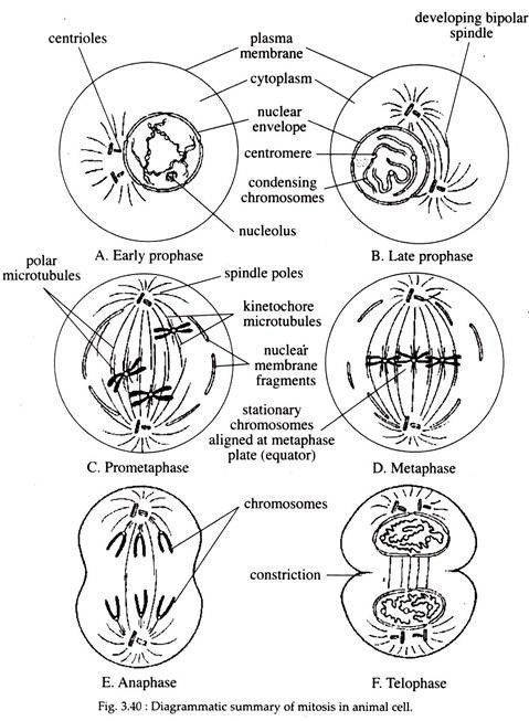
The process of mitosis is generally divided into four distinct stages — prophase, metaphase, anaphase, and telophase, each characterized by a particular series of events. However, such division of mitosis is arbitrary and is done only for the sake of discussion, since, mitosis is a continuous process and each stage represents a segment of that continuous process (Fig. 3.40).
Prophase:
The interphase nucleus contains extremely long, thin chromatin fibres spread throughout the entire nuclear compartment. The beginning of prophase is indicated by the condensation of these chromatin fibres by a process of folding.
ADVERTISEMENTS:
The prophase chromosomes appear to be composed of two coiled filaments, the chromatids, which are the results of the duplication of DNA during the S-phase. As prophase progresses, the chromatids become shorter and thicker in presence of an active enzyme, topoisomerase II.
The primary constriction (position of the centromere) contains the kinetochores become clearly visible and the evenly distributed chromosomes in the nuclear cavity approach the nuclear envelope resulting in an empty space at the centre of the nucleus.
In addition to the primary constrictions, secondary constrictions may be observed along some chromosomes. Other prophase changes include reduction in size of the nucleoli and their disintegration within the nucleoplasm.
The cell becomes spheroid, more retractile and viscous. At the end of the prophase, with rapid fragmentation and disappearance of the nuclear envelope, the nucleolar material is released into the cytoplasm.
ADVERTISEMENTS:
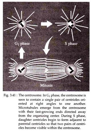
The most important cytoplasmic event that occurs in prophase is the formation of a microtubule – containing machine called the ‘mitotic spindle’ or ‘mitotic apparatus’. When an animal cell exits mitosis, it contains a single centrosome with two centrioles situated at right angles to one another.
During early prophase, microtubules appear in a ‘sun-burst’ arrangement or aster (Fig. 3.41), around each centrosome. This aster formation is followed by separation of the centrosomes from one another and their subsequent migration around the nucleus towards opposite sides of the cell.
As the pairs of centrioles separate, the microtubules stretching between them increase in number and elongate. Eventually, the two centrioles reach points opposite to one another, thus establishing the two poles of the mitotic spindle. Following mitosis, the two daughter cells get one centrosome each.
A number of animal cells (e.g., early mouse embryo) and plant cells lack centrioles. In such cells, during spindle formation, a clear zone appears around the nucleus. Microtubules rapidly accumulate in this zone during prophase, first in a dis-organised manner and then in a more oriented, spindle- shaped organisation.
In yeast and molds, the spindle forms entirely within the nucleus. Other cytoplasmic events occurring during prophase include extensive fragmentation of Golgi complex and ER to form large numbers of cytoplasmic vesicles that become dispersed throughout the cytoplasm. Other membranous organelles like mitochondria, lysosomes, peroxisomes, chloroplasts of plant cell, however, traverse mitosis relatively intact.
Metaphase:
The transition period between prophase and metaphase is called prometaphase. This is a short phase during which the dissolution of the nuclear envelope is completed, and the condensed chromosomes are scattered throughout the space that was the nuclear region.
At the beginning of prometaphase, the microtubules of the spindle penetrate into this space, so the free ends of some of the microtubules are ‘captured’ by a kinetochore and as a result the latter becomes firmly attached to the chromosome.
ADVERTISEMENTS:
Observations of living cells indicate that the attached chromosomes move, but not directly to the center of the spindle. Rather they oscillate back and forth along the chromosomal microtubules. It is suggested that the longer microtubules exert greater tension than the shorter micro-tubules.
As a result, the chromosomes tend to be pulled towards the pole from which the longer microtubule has extended. As the chromosomes move towards the center of the mitotic spindle, midway between the poles, the longer microtubules are shortened as a result of disassembly. The shorter microtubules on the other hand are elongated by the addition of tubulin subunits.
Eventually, each chromosome is moved into position along a plane at the center of the spindle, so that the microtubules from each pole are equivalent in length. Once all the chromosomes have become aligned at the spindle equator, with one chromatid of each chromosome connected to one pole and its sister chromatid connected to the opposite pole, the cell reaches the stage of metaphase.
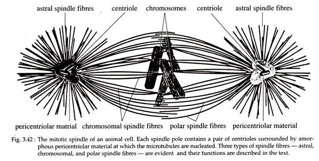
The plane of alignment of the chromosomes at metaphase is referred to as equatorial plate or metaphase plate. Functionally, the microtubules of the metaphase spindle can be divided into three groups (Fig. 3.42).
i. Astral Microtubules (Fibres):
They radiate out from the centrosome into the region outside the body of the spindle and help to keep the spindle in position.
ii. Chromosomal Microtubules (Fibres):
They extend from the centrosome to the kinetochores of the chromosomes and are required for the movement of chromosome towards the poles during anaphase.
iii. Polar or Inter-polar or Continuous Microtubules (Fibres):
They extend from the centrosome past the chromosomes and thus form a structural basket that maintains the integrity of the spindle (Fig. 3.42). In animal cells and some lower plant cells the spindle has centrioles and asters and is referred to as astral or amphiastral mitosis.
Mitosis in which centrioles and asters are absent is called anastral mitosis and is found in higher plants. However, centrioles and asters are not indispensable to the formation of the spindle.
Anaphase:
Anaphase begins with the sister chromatids suddenly splitting away from one another, due to release of a non-chromosomal protein ‘glue’ into the cytoplasm. All the chromosomes of the metaphase plate are split in synchrony and the chromatids (now referred to as chromosomes, since they are no longer attached to their duplicates) begin their pole-ward migration (Fig. 3.40).
This movement of each chromosome towards a pole is accompanied by the shortening of the microtubules attached to its kinetochore. During such movement the centrosome is seen at the leading edge With the arms of the chromosome trailing behind and thus the chromosome may assume the shape of a V with equal arms (if it is metacentric), or L with unequal arms (if it is sub-metacentric).
Some of these stretched spindle fibres now constitute the so-called inter-zonal fibres that appear between the pole-ward moving daughter chromosomes.
The movement of chromosomes towards the poles is referred to as anaphase A. The other movement that occurs simultaneously is referred to as anaphase B, in which the two spindle poles move further apart making the spindle elongated.
The latter movement is accompanied by net addition of tubulin sub- units to the plus ends (fast growing ends) of the polar microtubules. Thus, subunits can be added to polar or continuous microtubules and removed from chromosomal microtubules at the same time in different regions of the same cell. The movement of chromosomes during anaphase is very slow and about 1 µm per minute.
Telophase:
As the daughter chromosomes reach near their respective poles, telophase starts. During this stage, the chromosomes start to unfold and become less and less condensed by a process that recapitulates prophase but in reverse direction.
The chromosomes gather into masses of chromatin that become surrounded by discontinuous segments of nuclear envelope made by the ER (Fig. 3.40). Such segments then fuse to form two complete nuclear envelopes of the daughter nuclei. Finally, the nucleoli reappear at the sites of the nucleolar organizers.
Cytokinesis:
In animal cells, simultaneous with the formation of nuclear envelope, the cytoplasm constricts in the equatorial region. This constriction is accentuated and deepened until the cell cleaves into two. During cytokinesis, the cytoplasmic components, including mitochondria and Golgi complex are distributed between the two daughter cells (Fig. 3.40).
Cytokinesis in plant cells start with the formation of the phragmoplast that comprises the inter-zonal microtubules and Golgi vesicles. These structures are gradually transformed into the cell plate, which separate the territories of the daughter cells.
Within the cell plate the primary cell wall is produced mainly by pectin, which is contained in Golgi vesicles. Later on, micro-fibrils of cellulose are laid down in a semi-crystalline lattice on the two surfaces of the facing daughter cells.
Significances of Mitosis:
i. Mitosis helps the cell in maintaining proper size and equilibrium in the amount of DNA and RNA in the cell.
ii. The growth of multicellular organism is due to mitosis. After particular size has been reached, the cell divides to restore the nucleoplasmic index. Thus, growth takes place by an increase in the number of cells, rather than by increase in the size of the cells.
iii. The old, decaying and dead cells of the body are replaced by the process of mitosis. For example, dead cells of upper layer of epidermis, cells lining the gut and red blood cells, are constantly being replaced by mitosis.
iv. In certain organisms, mitosis is one of the means of sexual reproduction.
v. In multicellular organisms, gonads and sex cells depend on mitosis for increase in their number.


