In this essay we will discuss about:- 1. External Features of Earthworm 2. Transverse Section of Earthworm 3. Locomotion 4. Digestive System 5. Feeding and Digestion 6. Respiratory Organs 7. Blood Vascular System 8. Excretory System 9. Nervous System 10. Reproductive System 11. Reproduction.
Contents:
- Essay on the External Features of Earthworm
- Essay on the Transverse Section of Earthworm
- Essay on the Locomotion in Earthworm
- Essay on the Digestive System of Earthworm
- Essay on the Feeding and Digestion in Earthworm
- Essay on the Respiratory Organs of Earthworm
- Essay on the Blood Vascular System of Earthworm
- Essay on the Excretory System of Earthworm
- Essay on the Nervous System of Earthworm
- Essay on the Reproductive System of Earthworm
- Essay on the Reproduction in Earthworm
Essay # 1. External Features of Earthworm:
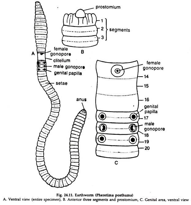
1. The body of the earthworm is long, narrow, almost cylindrical and slightly narrows down at both the ends. The anterior end is pointed, while the posterior end is more or less blunt.
ADVERTISEMENTS:
2. A full grown worm is about 150 mm long and 3 to 5 mm in diameter in the broadest portion of the body (Fig. 24.11A).
3. The dorsal surface is brown while the ventral surface is dull grey. A dark median line runs along the dorsal surface throughout the length of the body.
4. The surface bears a series of narrow ring-like grooves, and the body is divided into about 100 to 200 small segments or metameres.
5. The external segmentation corresponds to internal segmentation and the body is separated internally to a number of compartments.
ADVERTISEMENTS:
6. The mouth is a slightly ventral, crescent-shaped opening at the anterior end.
7. A small, fleshy lobe, the prostomium (Fig. 24.11B) is situated on the dorsal surface of the first or the buccal segment and overhangs the mouth.
8. At a distance of about 20 mm posterior to the anterior end, a prominent band known as clitellum is present.
9. The clitellum includes three segments 14 to 16, and divides the body into three distinct regions—the anterior or the preclitellar, the middle clitellar and the posterior post-clitellar regions.
ADVERTISEMENTS:
10. A median round aperture, the female genital pore is present on the ventral surface of the 14 segment (Fig. 24.11C).
11. A pair of crescentic apertures on raised papillae, the male genital pores are present on the ventral surface of the 18 segment.
12. In front and behind the male pores, on the 17 and 19 segments, a pair of papillae, the genital papillae are present in each segment. The ventral surface of the 17 to 19 segments is called the genital area.
13. Spermathecal pores or openings of the spermathecae are four pairs, placed ventrolaterally in the inter-segmental grooves between the 5 and 6, 6 and 7, 7 and 8 and 8 and 9 segments.
14. At about the middle of each segment except the first, the last, and the clitellar segments, chitinous needle-like structures called setae are arranged in a circle. The setae help in locomotion.
15. The dorsal pores are minute openings in the mid-dorsal line of inter-segmental grooves. Starting from the groove between the 12 and 13 segments and except the last groove, they are present in all the inter-segmental grooves.
16. The anus is a slit-like opening at the tip of the last segment.
17. The nephridiopores are minute, and scattered irregularly all over the skin except that of the first two segments.
Essay # 2. Transverse Section of Earthworm:
ADVERTISEMENTS:
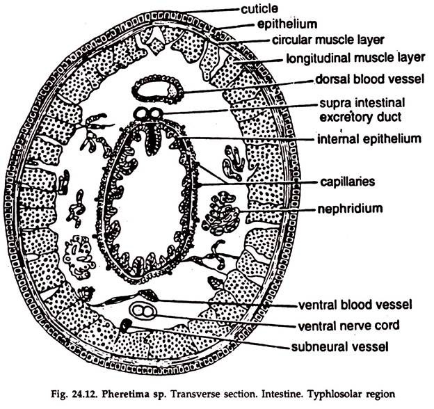
1. The outermost layer is a thin, porous cuticle secreted by the underlying epidermis (Fig. 24.12).
2. The epidermis is a single cell layer thick and consists of two kinds of cells, the large glandular cells with eccentric nucleus, and columnar cells placed between the glandular cells. A few cells functioning as sense cells are also present.
3. Dermis absent.
4. Two layers of muscles are present.
The outer circular muscular layer is narrow and continuous. The inner longitudinal muscular layer is thick and consists of muscle fibres arranged in narrow bands.
5. Other muscles absent.
6. Parietal layer. A single layer of coelomic epithelium forms the innermost layer of the body wall lining the coelomic cavity.
7. A distinct coelom is present. One dorsal, one ventral, one sub-neural blood vessel, two supraintestinal excretory ducts and one ventral nerve cord are present in the coelom. Coelom is divided into a number of compartments.
8. Parapodia absent. Chitinoid setae embedded in the body wall may be found.
9. The gut-wall consists of:
a. A visceral layer of coelomic epithelium.
b. Two layers of muscles — outer longitudinal and inner circular.
c. Enteric epithelium consists of a single layer of cells, some of which are glandular and others are absorptive. It is thrown into folds. A large median fold known as typhlosole in the region extending from 26 to 95 segments before anus, hangs in the lumen from the dorsal wall.
Essay # 3. Locomotion in Earthworm:
The locomotory structures are setae, circular muscle fibres arranged in the form of a ring, longitudinal muscle fibres in bands and the coelomic fluid.
Setae:
The setae are arranged in the form of a ring in all segments, except the first, last and clitellar segments.
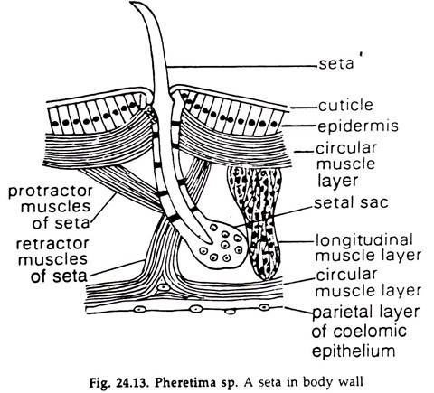
1. A seta is a narrow, elongated, chitinous structure hardened by sclerotised protein.
2. It is light yellow; the shape is in the form of a ‘f’ (Fig. 24.13), with a distal pointed end and a blunt proximal end embedded in setigerous sac in epidermal pit. A swelling, nodule, may be present in the middle.
Mechanism of Locomotion:
1. The setae of the posterior part of the body are fixed to the substratum; contraction of the circular muscles put pressure on the coelomic fluid and forces the longitudinal muscles to stretch and the anterior part of the body elongates.
2. In the next step, the setae of the anterior part are anchored; those of the posterior part released; the contraction of the longitudinal muscles increases the coelomic pressure, the circular muscles are stretched, leading to the shortening of the body and the earthworm moves forward.
Essay # 4. Digestive System of Earthworm:
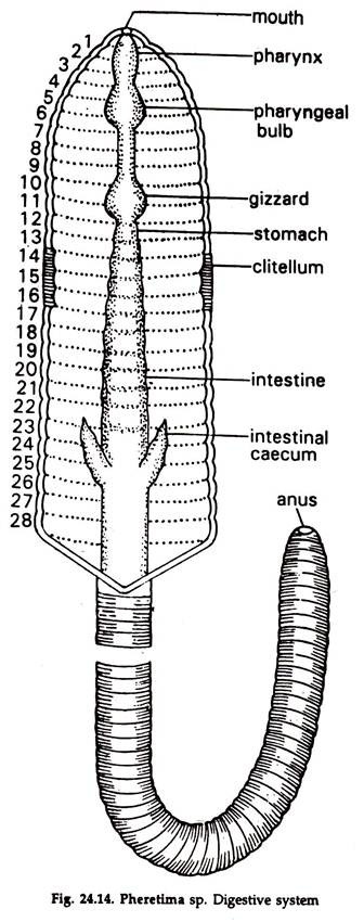
The alimentary canal is a straight tube, starting at the mouth and ending in the anus (Fig. 24.14).
Mouth:
The ventral mouth is crescent- shaped, situated in the peristomium. It is followed by a small buccal cavity, opening into the pharynx.
Pharynx:
The thick-walled, pear-shaped, muscular structure in the 3-4 segments. A pharyngeal bulb is present on the dorsal wall.
Oesophagus:
The muscular tube, extending from 5-8 segment and connects the pharynx with the stomach. The posterior part of the oesophagus is thick-walled, muscular with an inner, cuticular lining, oval in shape and known as gizzard.
Gizzard:
The food matters are grind to fine particles in it. It is a hard, muscular structure lined internally by a tough cuticle.
Stomach:
It is highly vascular, glandular, lies in 9-14 segments and continues posteriorly as intestine.
Intestine:
A thin walled, somewhat wide tube, running up to the anus in the last segment.
Intestinal coeca:
A pair of small, conical outgrowths, directed anteriorly, project from the sides of the intestine in the 26 segment.
Typhlosole:
A fold of the dorsal intestinal wall in the 26-95 segments, hangs downward in the lumen of the intestine (Fig. 24.12).
The intestinal wall is made of an outer peritoneal epithelium, longitudinal muscle fibres, circular muscle fibres and an inner epithelium of ciliated and glandular cells.
Essay # 5. Feeding and Digestion in Earthworm:
Earthworm obtains organic materials from the soil it swallow, rejecting the rest as cast. It also feeds upon decaying matters, small dead animals, etc. The mouth and pharynx help in feeding. In the pharynx mucin and proteolytic enzymes are secreted.
Mucin lubricates the food and digestion of protein starts. The gizzard grinds the food to fine particles and digestion is completed in the stomach with the help of enzymes secreted there. Absorption occurs in the intestine.
Essay # 6. Respiratory Organs of Earthworm:
Definite respiratory structures are lacking. The highly vascularized skin, kept moist by the secretion of epidermal glands and coelomic fluid coming out through dorsal pores, serves the functions of respiratory membrane.
Essay # 7. Blood Vascular System of Earthworm:
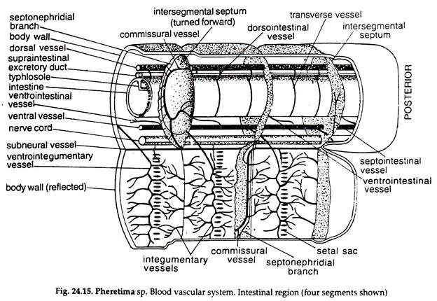
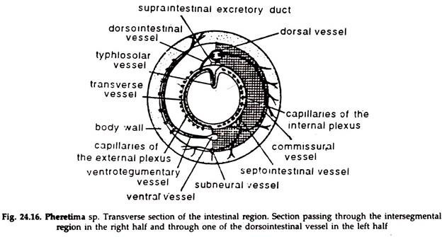
The blood of Pheretima is red due to the presence of haemoglobin dissolved in plasma. The corpuscles are colourless and nucleated. An elaborate system of blood vessels is found in earthworm. The arrangement of blood vessels is different in the intestinal region, i.e. posterior to the 13 segment, from that of the anterior region.
Blood Vessels in the Intestinal Region (Figs. 24.15, 24.16):
Dorsal blood vessel:
It runs along the mid-dorsal line between the gut and the body wall. It has a thick wall and contracts rhythmically. A pair of valves are present in the lumen of a vessel in each segment, to prevent backflow of blood.
It is a collecting vessel and receives blood from:
a. Two pairs of dorsointestinal vessels from the two ring-like vessels surrounding the intestine in each segment, bringing blood from the intestine.
b. A pair of commissural vessels from the posterior face of each septum receiving capillaries from the nephridia and body wall. The dorsal vessel drives the collected blood forward.
Ventral blood vessel:
It runs along the mid-ventral line just below the gut. It has no valves and acts as a distributing vessel by driving blood backward.
It sends the following vessels:
a. A pair of ventro-integumentary in each segment, which piercing the septum posteriorly, send blood to the body wall, septal and integumentary nephridia.
b. A median ventrointestinal, to the ventral wall of the intestine in each segment.
Sub-neural blood vessel:
It runs along the mid-ventral line below the ventral nerve cord. It extends from the posterior end of the body up to the 14 segment.
It is a collecting vessel and receives blood from:
a. A pair of small vessels in each segment bringing blood from the ventral region of the body wall.
b. The sub-neural vessel is connected with the dorsal vessel through a pair of commissurals in each septum.
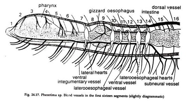
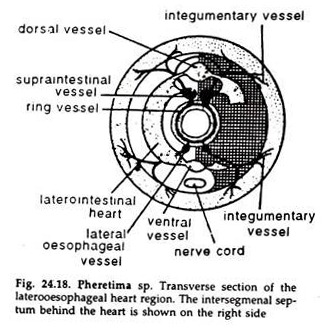
Blood Vessels in the Anterior Region (Fig. 24.17, 24.18):
The dorsal vessel runs in the usual position but acts as a distributing vessel, supplying blood to the pharynx and gizzard by small vessels and sending rest of the blood to the ventral vessel through the hearts.
The ventral vessel distributes blood in the usual fashion by a pair of ventro-integumentary vessels in each segment.
Hearts:
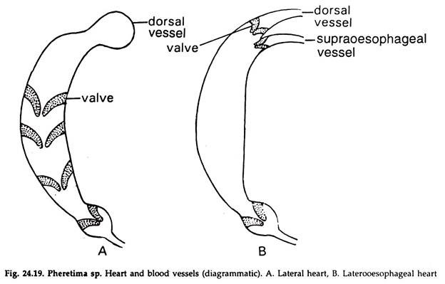
In the 7, 9, 12 and 13 segments, the dorsal vessel communicates with the ventral vessel through four pairs of pulsatile loops known as hearts. The first two pairs are called lateral hearts and the last two pairs latero-oesophageal hearts (Fig. 24.19).
The sub-neural vessel bends upward, bifurcates to two laterao-oesophageals and run by the sides of the gut up to the anterior end and act as collecting vessels.
In the region of 9 to 13 segments a short longitudinal vessel, the supraoesophageal vessel is present dorsal to the oesophagus. The latero-oesophageals are connected with the supraoesophageal by two pairs of loops in the 10 and 11 segments. These are called anterior loops. Blood from latero-oesophageals pass to the supraoesophageal through these anterior loops.
In the region of 12 and 13 segments the supraoesophageal is connected with the posterior two pairs of hearts through small vessels. The supraoesophageal and the dorsal vessel together communicate with the ventral vessel through the posterior two pairs of hearts and blood from the former passes to the latter through them.
Essay # 8. Excretory System of Earthworm:
Excretion or ‘removal of nitrogenous wastes is carried by nephridia. Nephridia are of 3 types. The septal and pharyngeal nephridia are ‘enteronephric’, but the integumentary nephridia are ‘ectonephric’.
Septal nephridia:
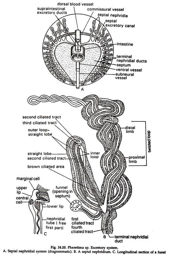
1. The septal nephridia are about 80-100 in each segment (Fig. 24.20A) posterior to the 15. The body of the nephridium consists of a short straight lobe and a long, spirally twisted loop (Fig. 24.20 B). The twisted loop is more than double the length of the straight lobe and is made up of two limbs; a proximal and a distal, the two spirally twisting each other.
The straight lobe continues into distal limb of the twisted loop at the base of the nephridium. The proximal limb of the twisted loop receives the ciliated ductule from the funnel and also gives out the terminal nephridial duct.
2. The body consists of a much coiled ciliated tubule richly supplied with blood.
3. The funnel bears a ciliated slit-like opening, the nephrostome, by means of which the nephridium is communicated with ‘he coelom. The funnel is lodged in the same segment in which the body of the nephridium lies. A narrow ciliated ductule connects the funnel with the body of the nephridium.
4. The terminal duct is a narrow continuation of the main body and the ducts of all the nephridia on half of each face of a septum unite to form a transverse duct, the septal excretory canal, which opens into the supraintestinal excretory duct of the side. A pair of supraintestinal excretory ducts are present on the dorsal surface of the gut and these ducts open in the gut by short vertical ducts near each inter-segmental partition. (Fig. 24.20A).
5. Wastes, collected by the nephridia are discharged into the’ lumen of the intestine and pass to the exterior with the excreta. As the nephridia open into the gut, they are called enteronephric nephridia.
Integumentary nephridia:
Smallest nephridia attached to the body wall and occur in large numbers in each segment, excepting the first two. Nephrostome absent, nephridiopore present. They are 200-500 in number in each segment, excepting the 14, 15 and 16, where they are numerous. All of them open directly to the exterior and termed ectonephric nephridia.
Pharyngeal nephridia:
Occur in large numbers in 4 to 6 segments as paired tufts associated with blood glands. Nephrostome absent. Terminal ducts unite to form a common duct and a pair of such ducts are present in each segment. These three pairs of ducts run forward in the fourth, fifth and sixth segments. The ducts of those in the 4 and 5 segments open in the lumen of the pharynx but of those in the 6 segment open in the buccal cavity.
Essay # 9. Nervous System of Earthworm:
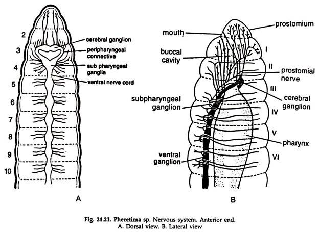
Nervous system is well-developed and includes central, peripheral and visceral nerves (Fig. 24.21).
Central Nervous System:
Cerebral ganglia or brain, peripharyngal connectives and the ventral nerve cord constitute the central nervous system.
1. Nerve ring. Situated at the anterior end of the body and vertical in orientation. The pharynx passes through it.
a. Supraoesophageal or cerebral ganglia. Two in number, fused to form the dorsal part of the ring or so-called brain.
b. Peripharyngeal connectives. Two, constitute the lateral sides of the ring around pharynx and connect the brain with the sub-pharyngeal ganglia.
c. Sub-pharyngeal ganglia. Two, fused to form the ventral portion of the nerve ring.
2. Ventral nerve cord. It is solid, consists of two nerves, right and left, enclosed in a sheath. It runs backward from sub-pharyngeal ganglia to the posterior end of the body, ventral to the ventral blood vessel. A ganglion in the ventral nerve cord is formed by the fusion of two ganglia.
Peripheral Nervous System:
1. Nerves emanating directly from the brain and ventral nerve coed are included in the peripheral nervous system.
2. Nerves from the brain innervate the prostomial region. A series of stomatogastric nerves from the peripharyngeal connectives pass to the pharynx and adjacent parts of the alimentary canal. The sub-pharyngeal ganglia send nerves to 2-4 segments.
3. Two pairs of nerves from each ganglion of the ventral nerve cord innervate the structures present in the segment.
Receptors and Sense Organs in Earthworm:
Three types of receptors have been identified in earthworm. A sensory fibre connects the base of each cells of the receptor to the central nervous system:
1. Epidermal receptors:
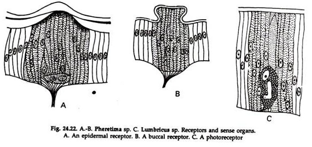
Long, slender cells, terminating in small, hair-like processes; tactile in function, are located in groups in the epidermis (Fig. 24.22A).
2. Photoreceptors:
They are present in the epidermis of the dorsal and dorsolateral sides; numerous at the anterior end; decreasing in number posteriorly. Each photoreceptor is a single cell (Fig. 24.22C) with a nucleus and a small transparent rod, the optic organelle (lens).
3. Buccal receptors:
The receptors for chemicals, smell and taste are called buccal receptors. They are groups of cells, present in large numbers in the wall of the buccal cavity. At the distal end the cells are broad and sensory hairs are better developed (Fig. 24.22B).
Essay # 10. Reproductive System of Earthworm:
Earthworm is hermaphrodite but auto-fertilization does not take place.
Male Reproductive System:
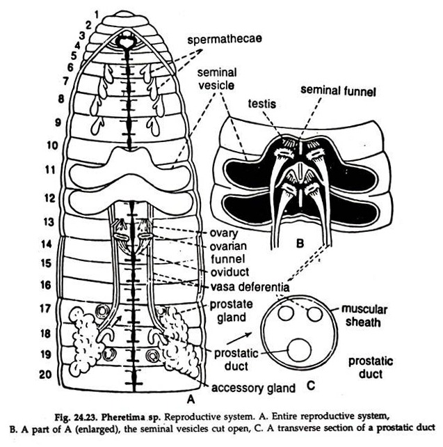
1. The male reproductive organs consist of two pairs of testes, two pairs of vasa deferentia with seminal funnels, two pairs of seminal vesicles, a pair of prostate glands with ducts and a pair of genital apertures (Fig. 24. 23A, B).
2. The testes are two pairs. They are small, white bodies composed of five to eight digitate processes coming out from a narrow base and are placed close to the middle line in the 10 and 11 segments.
3. A ciliated funnel, the seminal funnel is present just behind each testis and two pairs of funnels are present.
4. Each pair of testes and the respective seminal funnels are enclosed in a bilobed testis sac which continues behind into a seminal vesicle.
5. Each funnel leads posteriorly into a vas deferens and the two vasa deferentia of the side run backward up to the 18 segment.
6. A pair of white prostate glands, irregular in outline is present in the 16 to 22 segmental regions.
From each gland arises a duct, which is joined by the two vasa deferentia of the side and the three ducts are surrounded by a thick muscular sheath. This common structure is known as the prostatic duct. (Fig. 24.23C). All the three ducts, however, rim separately all along, and also open to the exterior separately through the male genital aperture situated on the ventral surface of the 18 segment.
7. Spermatozoa budded in the testes mature in the seminal vesicles and pass to the exterior through the vasa deferentia.
Female Reproductive System:
1. The female reproductive organs consist of a pair of ovaries, a pair of oviducts, a female gonopore and four pairs of spermathecae. (Fig. 24.23A).
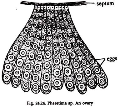
2. Ovaries are white bodies attached to the posterior face of the septum between the 12 and 13 segments. They resemble the testes very closely excepting the distal digitate processes, which are rather long. (Fig. 24.24).
3. Just behind each ovary is a ciliated funnel, the oviducal funnel, which ends in the oviduct. The two oviducts open externally by a common aperture placed on the ventral surface of the 14 segment.
4. Ova budded in the ovaries are shed in the coelom and being collected by the oviducal funnels pass to the exterior through the oviducts.
5. Spermatheca is a flask-shaped sac with an anterior blind diverticulum and opens to the exterior by a narrow duct. Sperms received from other partner during mating are stored up temporarily in these diverticula. Four pairs of spermathecae, one pair in each of the 6, 7, 8 and 9 segments are present.
Essay # 11. Reproduction in Earthworm:
Earthworm reproduces in monsoon through cross fertilization.
Copulation, Cocoon formation and Fertilization
Copulation:
During mating two individuals place themselves against each other in a head-to- tail position. The male genital aperture of one is placed against the spermathecal pore of the other. A mutual exchange of sperms and a certain amount of prostatic fluid take place.
Cocoon formation and fertilization:
Slime is secreted by the mucous glands in the clitellum, which forms a membranous .girdle enclosing both the partners. The earthworms start pulling them out of the girdle. While passing through the girdle, ova are discharged into it.
Sperms are discharged from the spermathecae into the girdle when the anterior ends of the animals pass through it. With the moving out of the animals, the ends of the elastic girdle close and a cocoon is formed. Fertilization and development take place inside the cocoon. Development is direct.
