In this article we will discuss about the development of scoliodon.
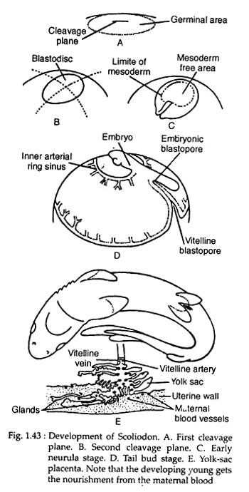
Scoliodon is viviparous and gives birth to living youngs. Fertilisation is internal. It is claimed that during copulation the claspers are introduced into the cloacal aperture of the female for the transmission of spermatozoa. The egg of Scoliodon is strongly telolecithal. The cleavage is restricted to a small germinal disc which floats on top of the yolk mass (Fig. 1.43A).
Cleavages are meroblastic and the first cleavage plane is simply a furrow in the surface of the germinal disc. The second cleavage occurs at right angle to the first one (Fig. T.43B). After this stage, the cleavage plane is irregular. By this way of cleavage, a blastodisc is separated from a layer of periblast cells.
ADVERTISEMENTS:
The periblast cells form a syncitial layer over the yolk mass. A blastocoel is formed between the blastodisc and the central region of the periblast. With the expansion of the blastocoel the blastodisc becomes multilayered.
The posterior region of the blastodisc grows faster than the other regions and is raised from the yolk mass to become a double-layered germ ring. This germ ring is raised posteriorly to form a blastopore (Fig. 1.43D).
At this stage an upper layer (epiblast) and a lower hypoblast is separated. The blastocoel lies between the ectoderm (epiblast) and the inner hypoblast (mesendoderm). The process of gastrulation actually starts with the formation of the dorsal lip of the blastopore. The endoderm is differentiated by cellular proliferation from the underside of the hypoblast. The mesoderm produces a prechordal plate.
As the embryo elongates along the anteroposterior axis, a head fold is marked off from the blastodisc and it becomes raised to form the neural folds. The body of the embryo is constricted from the blastodisc and the tail fold is differentiated and extended backward. The blastopore is separated into two (embryonic and vitelline blastopores) by the fusion of the edges of the yolk sac.
ADVERTISEMENTS:
The circulatory system appears in the mesoderm in the form of blood islets which unite to form vascular networks. As the embryo grows, mouth is formed from the stomodeal invagination and anus is formed anew from the proctodeal invagination because the embryonic blastopore becomes closed.
The developing embryo is provided with a tubular yolk stalk. This yolk stalk connects the intestine of the embryo with the yolk sac (Fig. 1.43E). The yolk sac contains yolk material which provides nutrition for the developing embryo. When the yolk is fully exhausted the yolk sac becomes folded and becomes anchored with the uterine wall of the mother as the yolk-sac placenta.
With the formation of placenta, the yolk stalk is lost and the blood vessels of the yolk stalk form the placental cord. The placental cord connects the embryo with the yolk-sac placenta. The placental cord develops many finger-like processes called appendicula.
Each appendicularium is made up of a central core of loose connective tissue surrounded by several layers of epithelial cells. The distribution of the appendicula in the placental cord varies in different regions. The appendicula help in the absorption of nutrients secreted from the uterine wall of the mother.
