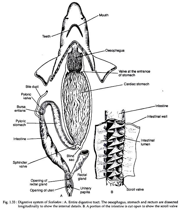
The digestive system of scoliodon consists of the alimentary canal and the digestive glands. The alimentary canal starts with the mouth and terminates in the anus. The mouth leads into a spacious buccal cavity which is lined with mucous membrane.
The floor of the buccal cavity becomes folded to form a non-muscular and non-glandular tongue. The mucous membrane is very thick and rough due to the presence of dermal denticles or teeth. The teeth are very sharp and are obliquely placed. The teeth are homodont (i.e., the teeth are similar in shape) and possesses several sets of teeth functioning in succession.
The buccal cavity leads into pharynx. On either side of the pharynx there lie the internal openings of the spiracles and five branchial clefts. The mucous membrane of the pharyngeal wall contains numerous dermal denticles. The pharynx leads into a narrow oesophagus. The inner mucous membrane of the pharynx is raised to form longitudinal folds.
The oesophagus dilates posteriorly to form a large stomach. The stomach is highly muscular and is bent on itself to form a J-shaped configuration. The long limb of the stomach is continuous with the oesophagus and the shorter one passes into the intestine. The entrance of the oesophagus into the stomach is provided with a crescentic fold which serves as the valve.
ADVERTISEMENTS:
The long anterior limb is called the cardiac stomach and the short posterior limb is designated as the pyloric stomach. A small outgrowth, often called ‘blind sac’, is present at the junction of the cardiac and pyloric limbs. The inner lining of the cardiac stomach is folded longitudinally like that of oesophagus (Fig. 1.33A).
The internal lining of the pyloric stomach is mostly smooth though slight foldings are observed at the distal end. The pyloric valve, at the end of the pylorus, guards the entrance of it into a thick-walled small chamber called the bursa entiana.
The bursa entiana is immediately followed by wide tubular intestine which becomes narrowed posteriorly as the rectum. The rectum opens into the cloaca. A tubular caecal or rectal or digitiform gland opens into the rectum.
ADVERTISEMENTS:
The inner surface of the intestine becomes folded to form an anticlockwise spiral of approximately two and a half turns. This is called the scroll valve (Fig. 1.33B) which increases the absorptive surface of the intestine and also checks the rapid flow of digested food through the intestine.
The major digestive gland is the liver which is a massive yellowish gland and consists of two lobes. The lobes are united anteriorly. A thin-walled V-shaped gallbladder is present in the anterior part of the right lobe of liver. The bile duct receives few smaller ducts from the two lobes of the liver and opens into the anterior end of intestine, near the commencement of the scroll valve.
The pancreas is a pale compact irregular body and consists of a dorsal lobe situated parallel to the posterior part of cardiac stomach and a ventral lobe which remains closely attached to the pyloric stomach. The pancreatic juice is poured into the intestine by pancreatic duct situated opposite to the aperture of the bile duct.
The functional significance of rectal gland is not properly known. The rectal gland has a central cavity lined with cuboidal cells. It is highly vascular and composed of lymphoid tissue. It discharges a fluid into the lumen of the intestine but its actual role is not known.
ADVERTISEMENTS:
The buccal cavity possesses no such glands that can be compared with the salivary glands of higher vertebrates. The spleen is located dorsal to the distal end of the body of the stomach. The spleen is functionally associated with the circulatory system, but remains morphologically connected with the alimentary canal.
