The nervous system of Channa Punctatus comprises of the central nervous system, peripheral nervous system and the sympathetic nervous system.
Central nervous system:
This system includes the brain and the spinal cord.
Brain:
ADVERTISEMENTS:
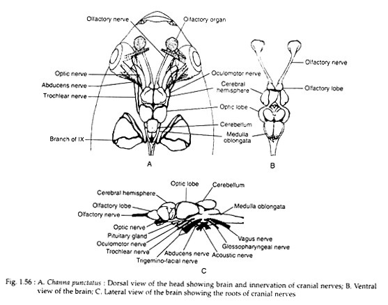
Brain (Fig. 1.56A-C) in Channa punctatus is well developed and remains covered with good amount of adipose tissue. The olfactory lobes and cerebral hemispheres are distinct. The moderately developed oblong olfactory lobes, as usual, occupy the anterior part of the telencephalon.
Remarkably, the large cerebral hemispheres are characterised by two convolutions in each. Of these, the dorsal one is slightly larger than the anterolateral one. The large oval inferior lobes are located at the ventral aspect of diencephalon. The lobes encircle the infundibulum which at its apex bears the pituitary.
The reduced saccus vasculosus, extending backward from the infundibulum, is partly housed in the cleft of the adjoining inferior lobes. Moderately developed globular optic lobes form the widest part of the brain. These are followed by moderately developed cerebellum.
It is roughly rectangular in outline and the small auricular lobes are present at the lateral aspect of the cerebellum. The medulla oblongata is characterised by large which exist as paired humps over dorsal part of the former.
Cranial nerves:
Ten pairs of cranial nerves are discernible in Channa punctatus. The cranial nerves are read in two ways. In one way the nerves are called on the basis of numbers, like Ist, IInd, IIIrd … etc. In other way they are called by name. Here both number and names are mentioned.
I. Olfactory nerve (Fig. 1.56):
It arises from the anterior aspect of the olfactory lobe as a thick cylindrical cord. It then projects forward and outward to reach the base of the olfactory rosette emerging through the olfactory foramen. The nerve finally innervates the olfactory lamella.
ADVERTISEMENTS:
II. Optic nerve (Fig. 1.56A, C):
This thicker cylindrical nerve arises from the anteroventral part of the optic lobe and at the region of the optic chiasma, the nerve undergoes superficial longitudinal splittings. At this region the nerve of the left side passes over the right. Finally the nerve enters the optic capsule to innervate extensively the retina.
III. Oculomotor nerve (Fig. 1.56):
This cylindrical, moderately developed nerve arises from the anterolateral part of the medulla. Leaving the brain, the nerve runs straight forward and then splits into superior and inferior branches at the base of posterior eye muscles.
IV. Trochlear nerve (Fig. 1.56):
This nerve originates from the anterolateral part of the medulla oblongata. Its forward course is diverged slightly outward to reach the superior oblique muscle which it innervates.
VI. Abducens nerve (Fig. 1.57):
ADVERTISEMENTS:
It is the thinnest of the cranial nerves and arises from the anteroventral part of the medulla oblongata. Moving forward along the ventral plane of the brain, the nerve reaches the external rectus muscle to innervate it.
V & VII. Trigeminal and Facial nerves (Fig. 1.57A):
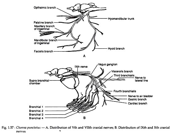
The trigeminal nerve arises from the anterolateral part of the medulla oblongata in the form of two laterally compressed broad bundle of nerves. Four more bundle of nerves arise from the posterior aspect of the trigeminal root which actually form the root of facial nerve.
Of these four bundles, three along with two of trigeminal, coalesce to form a complex mass. This mass further receives a short connective from the remaining facial nerve bundle (hyomandibular trunk) and thus a trigeminofacial ganglionic complex is formed. The ganglionic complex is located intra-cranially in the mesial aspect of the prootic bone.
Three main nerve trunks arise from the trigeminofacial ganglionic complex – the supraorbital, the infraorbital and the hyomandibular in addition to palatine nerve. The supraorbital and infraorbital group of nerves exit through the anterior opening of the prootic bone.
VIII. Acoustic nerve (Fig. 1.56C):
This short compressed nerve arises from the anterolateral part of the medulla. Root is located between the roots of facial and glossopharyngeal nerves. Soon after origin, the nerve courses downward and then trifurcates to innervate different parts of internal ear. The anterior branch further divides to produce four twigs.
One supplies the ampulla of the horizontal semicircular canal, two branches to the utriculus and the rest one to the ampulla of the anterior semicircular canal. The posterior branch bifurcates to innervate the ampulla of the posterior semicircular canal and lagena. The remaining branch (middle), projecting downward, ramifies largely to innervate the sacculus.
IX. Glossopharyngeal nerve (Fig. 1.56C, 1.57B):
This cylindrical nerve arises from the ventrolateral part of the medulla oblongata through a single root. Turning backward, the nerve comes out from the cranium through a fine foramen at the exoccipital. Outside the cranium the nerve follows a forward course and then creeps upward along the dorsal plane of the supra-branchial organ.
The ganglion (petrosal ganglion) is formed over this supra-branchial chamber which produces two branches — one anterior and the other lateral. The anterior branch innervates the wall of the supra-branchial chamber whereas the lateral branch descends along the lateral part of the first gill arch and finally terminates in the ventral branchial muscles.
Initially, as it reaches the first gill arch, it produces finer branches which innervate the lining membrane of the area concerned. While on the supra-branchial organ, a finer branch (pharyngeal branch) is further given off which moves backward to innervate the dorsal branchial muscle.
X. Vagus nerve (Fig. 1.57B, 1.58):
The vagus nerve arises by two roots from the lateral surface of the medulla oblongata, posterior to the origin of glossopharyngeal nerve. These two nerves soon coalesce to form a common nerve which runs posteriorly and ultimately comes out of the cranium through a foramen on the exoccipital.
Outside the cranium it forms a swelling (vagus ganglion) from which arises several nerves of variable magnitude. Form innervation point of view these nerves may be categorized into three groups: (a) ramus lateralis, (b) ramus visceralis, (c) ramus branchialis.
(a) Ramus lateralis:
These are the outermost group of nerves arising from the lateral most part of the vagus ganglion to innervate the sensory canals of the head and trunk region (lateral line). The main trunk of the lateralis is a broad flat nerve which, on leaving the vagus ganglion, enters deep into the body to reach the vertebral column.
The nerve runs straight backward up-to the caudal end adhering to the vertebral column throughout its length. On its way, the lateralis sends several subcutaneous branches which innervate different sections of truncal canal.
(b) Ramus visceralis:
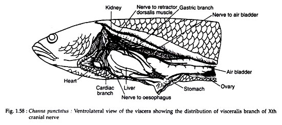
It arises as a single stout nerve from the vagus ganglion and on moving downward very soon divides to form three branches. The dorsal most branches splits into two nerves both of which innervate the retracto dorsalis muscle.
The middle branch enters deep into the visceral cavity and bifurcates to produce dorsal and ventral branches. The dorsal branch moves straight backward for quite a long distance along the ventral part of the kidney and finally innervates the air bladder.
The ventral branch very soon ramifies over the stomach and innervates it. The remaining ventral branch (cardiac branch) of the visceralis proper is much thicker initially but later bifurcates to form a well- developed branch that innervates the sphincter oesophagi muscle and a thinner branch which, on further ramification, innervates the heart.
(c) Ramus branchialis:
Altogether four branchial nerves (on each side) are present and innervate the dorsal and ventral branchial muscles, gill lamellae and the gill rakers. The first branchialis arises independently from the vagus ganglion and emerges through the cleft of levator muscles to become exposed over the roof of the branchial chamber.
Initially, this nerve produces a short branch (pharyngeal branch) which immediately ramifies to innervate the levator externus muscle and floor of the supra-branchial chamber.
The main trunk of the first branchialis moves outward and bifurcates to form a thinner pretrematic branch and a thicker posttrematic branch. The pretrematic branch runs along the base of the lamella of first gill arch and innervates it. The posttrematic branch moves downward along the ventral side of the second gill arch.
The second branchialis also arises independently from the vagus ganglion. A distinct nerve (pharyngeal branch) courses forward from the main trunk of the second branchialis, moves below the levator externus muscle and finally ramifies over the trans versus dorsalis anterior muscle to innervate it.
The second branchialis proper, like the first one, bifurcates at the dorsolateral part of the branchial basket to form pre- and posttrematic branches. The slender pretrematic branch innervates the gill lamella of the second gill arch, whereas the posttrematic branch proceeds along the ventral trough of the third gill arch.
The third and fourth branchialis arise through a common trunk from the base of the visceralis branch. These two nerves are comparatively thinner than the former two (first and second branchialis) and remain almost side by side for quite a considerable distance.
The finer pretrematic branch of the third branchialis branch, proceeds along the ventral trough of the fourth gill arch. Initially, while moving over the dorsal branchial muscles, the third branchialis innervates the obliquus dorsalis muscle by a fine pharyngeal branch.
The fourth branchialis, at the very onset, delivers a fine pretrematic branch which innervates the gill lamella of the fourth arch and then curves anteriorly along the posteroventral part of the branchial basket as a flat nerve. Main trunk of the fourth branchialis proceeds along the ventral trough of the fourth gill arch as the posttrematic branch.
Sense organs:
The main sense organs are the eyes, ears, olfactory organs, receptors for touch, and the lateral line sense organs.
(i) Eyes:
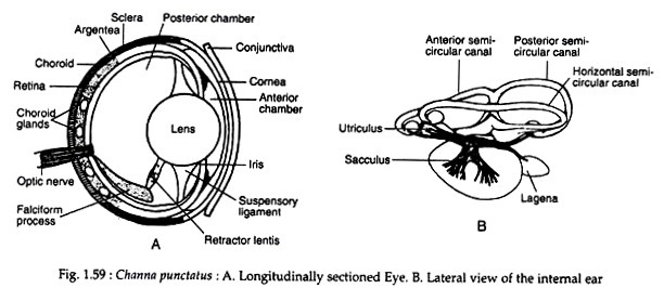
The paired eyes are the photoreceptors. The eyeball is three-layered. The outer or sclerotic layer contains cartilage, a median vascular choroid layer and an innermost photosensitive retinal layer. The choroid layer contains the choroid glands which surround the optic nerves. The eyes are devoid of eyelids. The cornea is flat.
The lens is globular and lies in apposition to the cornea. The anterior chamber of the eye is greatly reduced. Between the choroid and the sclerotic layers there is a silvery reflecting layer or argentea. There is no ciliary muscle but there is a peculiar falciform process in the posterior chamber of the eye which helps in changing the position of the lens (Fig. 1.59A).
The falciform process is the vascular fold of the choroid. This fold pierces the retinal layer near the entrance of the optic nerve and proceeds to the back of the lens. The back of the lens is attached to the falciform process by a retractor lentis muscle.
Each eye has different visual field and the vision is thus monocular. Accommodation is effected by shifting the position of the lens and not by changing the shape of the lens as observed in higher vertebrates.
(ii) Ear:
The ear is exclusively composed of internal ear or membranous labyrinth. The middle and external ears of higher vertebrates are absent. The membranous labyrinth is composed of an upper chamber called utriculus and a lower sacculus (Fig. 1.59B). Three semicircular canals open into the utriculus.
The sacculus is a sac-like structure whose floor gives rise to a lagena. The endolymph contains ear-stones or otoliths which touch the sensory hairs of both the chambers. The semicircular canals and the utriculus help the animal to maintain equilibrium. The sacculus and the lagena perceive the sound waves.
As there is no tympanum, the body surface transmits the vibration to the inner ear. The lateral line sense organ helps the fish to perceive low frequency vibrations in water. The ears thus constitute the audio-equilibrating sense organs. Although the organ of Corti (the sensory area responsible for hearing in higher vertebrates) is lacking, this fish can undoubtedly hear.
(iii) Lateral line sense organs:
The lateral line sense organs are well-developed in Lata. This lateral line system is composed of a large number of deep-seated sense organs (neuromasts). Each sense organ is housed in a pit and is communicated to the surrounding water by a pore. The lateral line sense organs are innervated by the lateral line branch of the vagus. This sensory system primarily helps the animal to perceive low frequency vibrations in water.
(iv) Sense organs for touch:
There is no such definite organ but the tactile cells on the lips and on the surface of the body serve the purpose.
(v) Olfactory sense organ:
The olfactory sense organ is represented by two blind nasal sacs. The olfactory cells lining the nasal sacs are sensitive to smell. The nasal sacs do not communicate to the buccal cavity but open to the exterior by openings. The nasal sacs are exclusively olfactory in function and do not play any role in respiratory system.


