The nervous system of Scoliodon includes:
(i) The central nervous system,
(ii) The peripheral nervous system, and
(iii) The autonomous nervous system.
ADVERTISEMENTS:
(i) Central nervous system:
The central nervous system consists of brain and the spinal cord.
(a) Brain:
Brain is highly organised and shows many advancements over that of the agnathans.
ADVERTISEMENTS:
The brain is divided into three primary parts:
(a) The forebrain or prosencephalon,
(b) The midbrain or mesencephalon
(c) The hindbrain or rhombencephalon.
ADVERTISEMENTS:
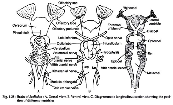
The forebrain consists of a massive undivided cerebral hemisphere. The cerebral hemisphere is relatively larger than that of other fishes. From the anterior end of cerebral hemisphere arise two stout olfactory peduncles; each terminates into a large bilobed olfactory lobe (Fig. 1.38A).
The olfactory lobes lie close to the olfactory capsules. Each olfactory nerve is composed of many bundles of nerve fibres. The surface of the cerebrum is smooth and the walls are thick. A small opening called the neuropore is present on the mid-ventral surface of the cerebrum. The posterior part of forebrain (diencephalon) is very short. The roof of the diencephalon is thin, non-nervous and contains the anterior choroid plexus.
The lateral walls of the diencephalon form two thickened bodies called thalami. A long and slender tube, the pineal organ or epiphysis cerebri projects from the roof of the diencephalon. The floor of the diencephalon (or hypothalamus) is well-formed. A hollow infundibulum is given off from the floor of the diencephalon.
The infundibulum is dilated to form two oval thick-walled bodies called lobi inferiores whose distal ends are produced into two thin-walled glandular sacs called sacci vasculosi. The lobi inferiores are the centres for gustation and smell.
The hypophysis is attached to the infundibulum. The optic chiasma lies in front of the infundibulum. The optic chiasma is formed by the decussation of the nerve fibres of two optic nerves (Fig. 1.38B).
The midbrain is large and consists of two round optic lobes. The optic lobes are situated behind the diencephalon. The floor and the side walls are relatively thicker. The midbrain is considered as the centre of coordination.
The hindbrain consists of a highly developed cerebellum and a medulla oblongata. The dorsal surface of the cerebellum produces many irregular convolutions. The cerebellum contains a small cavity. The cerebellum is also a centre of co-ordination. The cerebellum is divided into three lobes by two well-marked transverse furrows.
The medulla oblongata is triangular and the anterior end gives a pair of hollow corpora restiformia with trace of convolutions in adults. The medulla controls respiration. Two corpora restiformia are connected by the transverse nerve band. The roof of the medulla oblongata is non-nervous and bears the posterior choroid plexus. The hind- brain controls swimming movements.
ADVERTISEMENTS:
The ventricles of the brain are moderately developed (Fig. 1.38C). The cerebral hemispheres contain narrow lateral ventricle. The third ventricle is extended forward about half the length of the cerebral hemispheres. The floor of the fourth ventricle is very much thickened.
The fourth ventricle is large and extends dorsally into the cerebellum and is continuous behind with the cavity of the spinal cord. The iter (i.e., the communicating duct between the third and the fourth ventricles) is wider. Although the cerebrum is undivided, there are two lateral ventricles which are continued to the rhinocoels (cavity of the olfactory lobes).
(b) Spinal cord:
The spinal cord in Scoliodon shows definite advancement towards the plan of higher vertebrates. The grey matter is arranged into the dorsal and the ventral horns. The dorsal horns are united to form a single broad region; as a result the grey matter assumes a shape of an inverted ‘T’.
(ii) Peripheral nervous system:
The peripheral nervous system includes the cranial nerves and spinal nerves.
(a) Cranial nerves:
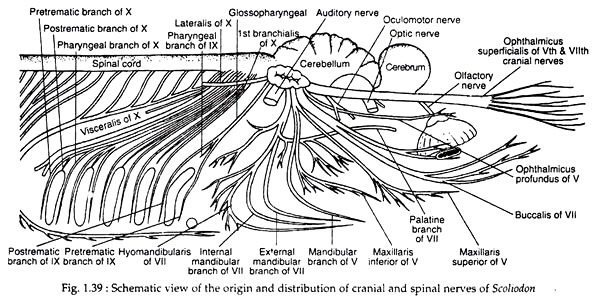
There are ten pairs of cranial nerves in all the fishes. The first pair of cranial nerves is the olfactory nerves which originate from the olfactory lobes and innervate the olfactory sacs. The terminal nerves are situated between the two olfactory lobes. These nerves emerge from the telencephalon and bear a ganglion called ganglion terminale near the origin.
These nerves supply the nasal septum and the external nostril. The second pair of cranial nerves is the optic nerves which, after the origin from the optic thalami, form the optic chiasma and supply the eyes. The third cranial nerve is called oculomotor nerve which originates from the ventral surface of the mesencephalon and supplies the anterior, superior and inferior recti and the inferior oblique muscles of each eye ball.
The fourth cranial nerve is called trochlear or pathetic nerve which arises from the dorsolateral surface of the midbrain and supplies the superior oblique eye muscle.
The fifth cranial nerve is the trigeminal which has three branches:
(1) Ophthalmicus superficial which supplies to the skin of the snout.
(2) The maxillaris which is divided into maxillaris superior supplying nerves to the skin of the upper jaw and maxillaris inferior innervating the posterior part of the upper jaw.
(3) The mandibularis innervating the muscles of the lower jaw. Another nerve called ophthalmicus pro-fundus becomes secondarily associated with the trigeminal to supply nerves to the eye ball and the dorsal surface of the snout (Fig. 1.39).
The sixth cranial nerve is the abducens which supplies the posterior rectus muscle of the eye ball. The seventh cranial nerve is known as facial which divides into two branches:
(1) The ophthalmicus superficialis branch like that of the fifth cranial nerve.
(2) A bundle of mixed nerves which subdivides into three routes:
(a) A ramus buccalis innervating the infraorbital canal of the snout.
(b) A ramus hyomandibularis supplying nerves to the lower jaw and throat.
(c) A ramus palatinus giving nerve supply to the roof of the buccal cavity and the pharynx. The eighth cranial nerve is called auditory which gives the vestibular and saccular branches to the internal ear.
The ninth cranial nerve is the glossopharyngeal which, in the region of the first gill cleft, divides into a small pretrematic nerve and a large posttrematic nerve. These nerves supply branches to the pharynx, pharyngeal muscles and the mucous membrane surrounding the first gill slit.
The tenth cranial nerve is the vagus which arises by multiple roots and gives off many branches.
The branches are:
(i) the brachial nerves supplying the gills,
(ii) the lateralis supplying the lateral line sense organs and gives numerous branches along its course.
(b) Spinal nerves:
The spinal nerves arise from the spinal cord. Each has one dorsal and one ventral root. The dorsal root bears a ganglionic swelling. After emerging out through the vertebral column, the dorsal and the ventral roots unite to form a common mixed nerve.
Each spinal nerve gives three branches, such as:
(a) Ramus dorsalis,
(b) Ramus ventralis, and
(c) Ramus communicans to join with the autonomous nervous system.
(iii) Autonomous nervous system:
This system is made up of a series of paired ganglia arranged irregularly on the dorsal wall of the kidney and the posterior cardinal sinuses. The gastric ganglion is the largest ganglion and sends nerves to the viscera. There is usually one ganglion in each segment. In Scoliodon the successive ganglia are not in distinct continuous chain.
Sense Organs:
The nervous system is associated with highly developed sense organs, viz., eyes, nose, ear and many others (Fig. 1.40).
(i) Eyes:
The eyes are built on the principle of a photographic camera. The eyeball is composed of three layers: the sclera, choroid and retina. The sclera is cartilaginous. The pupil is a vertical slit and it cannot be dilated or contracted. The retinal layer consists of rods and cones. A large number of guanine plates are present on the inner surface of the choroid layer which is called tapetum lucidum.
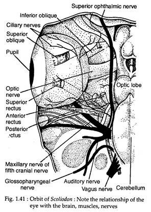
It acts as reflector. The crystalline lens is spherical. The lens is kept in position by suspensory ligament which extends from the margins of the lens to the ciliary processes. The ciliary processes are longitudinal folds of the choroid layer. The eye is kept in its position in the orbit by six extrinsic eye muscles. Besides the eye muscles there are an optic nerve and a cartilaginous optic pedicle (Fig. 1.41).
These eye muscles are attached with the eyeball in two groups. The first group includes:
(i) Superior rectus,
(ii) Inferior rectus,
(iii) Anterior rectus, and
(iv) Posterior rectus.
The second group includes:
(i) Superior oblique and
(ii) Inferior oblique.
The superior rectus muscle runs outwards and upwards and is inserted on the dorsal side of the eye ball. The inferior rectus muscle extends outwards and downwards to be inserted on the ventral side of the eyeball. The anterior rectus muscle runs outwards and downwards and is attached to the ventral surface of the eyeball.
The posterior rectus muscle runs backwards and is attached with the posterior surface of the eyeball. The superior oblique muscle is attached on the dorsal surface of the orbit just in front of the insertion region of the superior rectus muscle.
The inferior oblique muscle is attached to the ventral surface of the eyeball just in front of the inferior rectus muscle. The eyes of Scoliodon are quite prominent and are proportionately larger in size. The eyes are laterally placed and each eye has its own range of vision, i.e., monocular vision. Scoliodon is possibly colour-blind.
(ii) Olfactory organs:
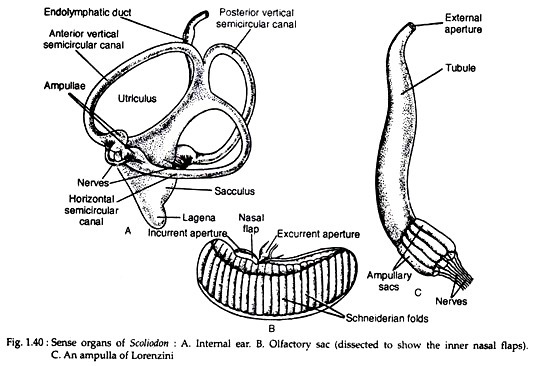
There are two blind sac-like olfactory organs situated in front of the mouth. Each olfactory sac is placed in a cartilaginous capsule and does not communicate with the buccal cavity. The mucous membrane of the olfactory sac is thrown into two series of folds called Schneiderian folds which are placed by the median raphe (see Fig. 1.40B).
The Schneiderian folds are composed of olfactory sense cells and supporting cells. Physiologically each nasal opening is divided by three muscular nasal valves into a median ex-current siphon and a lateral incurrent siphon.
(iii) Internal ear:
The internal ear or the membranous labyrinth of Scoliodon is a closed ectodermal sac. It consists of three semicircular canals and a central body differentiated into an anterior utriculus and a ventral sacculus (see Fig. 1.40A). The three semicircular canals are arranged in three planes of the body and open into the central body by both ends.
But one end of each canal dilates to form ampulla. The utriculus gives off an invagination called recessus utriculi beneath the ampullae of the anterior vertical and horizontal semicircular canals. The sacculus gives a posterior outgrowth called the lagena. The membranous labyrinth is placed in a cartilaginous auditory capsule.
The inner cavity is filled with a fluid called the endolymph into which many calcareous bodies (otoliths) are present. The space between the membranous labyrinth and the auditory capsule is filled with the perilymph. A long tube called the endo-lymphatic duct communicates the cavity of sacculus to the exterior through a small opening.
The membranous labyrinth of Scoliodon performs three functions:
(1) It helps in orientation in relation to gravity,
(2) It accelerates in changing the direction during swimming, and
(3) It helps in hearing.
The utriculus together with the semicircular canals is responsible for the orientation and acceleration, while the sacculus is meant for hearing. The internal ears are the statoacoustic organs in Scoliodon.
(iv) Neuromast organs:
These organs comprise of the lateral line receptors and the pit organs. The lateral line sense organs lie inside the lateral line canal. These sense organs are the epidermal derivatives and are called neuromasts which remain embedded in the wall of the lateral line canal. This canal communicates to the exterior by minute pores.
The pit organs occur on the lateral and dorsal sides of the head. They are actually ectodermal pits connected by groups of sense cells. The neuromast organs help to orient the body in relation to currents and waves and are designated as rheoreceptors.
(v) Ampullae of Lorenzini:
Innumerable pores on the dorsal and the ventral sides of the head lead into a long tube which terminates into radially arranged ampullary sacs (Fig. 1.40C). The ampullae lie in clusters and each of these consists of eight or nine chambers arranged radially around a central core called centrum.
Two types of cells are encountered in the ampullae—glandular cells and sensory cells. The ampullae get the names according to their location. These sense organs are the thermo receptors and are also the pressure receptors.


