In this article we will discuss about the exoskeleton and endoskeleton structure of scoliodon.
The exoskeleton includes predominantly the scales which are present all over the body. Endoskeleton comprises of the axial and appendicular skeleton.
Exoskeleton:
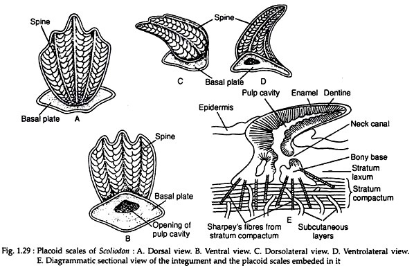
The entire surface of the scoliodon body is covered by oblique rows of placoid scales or odontoids (Fig. 1.29). Atypical placoid scale has a basal plate made up of calcified tissue which remains embedded in the skin and a backwardly directed spine projecting out of the skin.
The basal plate is held in the dermis by Sharpey’s fibre and other fibres (Fig. 1.29E). A perforation is present at the base of the spine which communicates with the pulp cavity of the basal plate. The spine is composed of dentine coated externally with enamel.
ADVERTISEMENTS:
The nature of enamel is controversial. It is called the fibro-dentine, because it is formed as the calcification of the fibrous material between the dentine and the enamel organ. The basal plates as well as the dentine of the spine are the derivatives of the mesoderm. The enamel is derived from the ectodermal enamel organ.
Endoskeleton:
Endoskeleton is composed exclusively of cartilage which may be hardened by calcification at places.
It can be described under two heads:
ADVERTISEMENTS:
(1) The axial skeleton comprising of the skull and the vertebral column.
(2) The appendicular skeleton consisting of the pectoral and pelvic girdles and the skeleton of the fins.
(1) Axial skeleton:
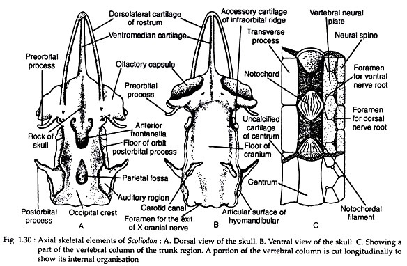
The skull is a simple cartilaginous casket. It consists of the cranium, four sense capsules enclosing the auditory and olfactory organs and the visceral skeleton which form the jaws and supports the pharynx along with the gills. These three components of the skull remain intimately fused with each other (Fig. 1.30A, B).
The cranium has four distinct regions:
(1) The occipital region forms the posterior portion of the skull and contains a large foramen magnum through which the spinal cord passes down. An occipital condyle is present on either side of the foramen magnum. Above this foramen there is a median ridge called the occipital crest.
(2) The auditory region is made of auditory capsules which remain firmly united with the cranium in an adult. An oval depression between the two capsules is called the parietal fossa. The roof of the cranium has two large fontanelles on the dorsal side.
(3) The middle region of the skull is composed of orbit. The margin between the roof of the cranium and the orbit is marked by the supraorbital ridge. A slender cartilage called pre-orbital process partly encircles the orbit. Similarly a post-orbital process emerges from the side forward along the upper margin of the orbit.
(4) The anterior or the ethmoidal region of the cranium is composed of olfactory capsules and rostrum. The two olfactory capsules are separated by internasal septum.
The visceral skeleton consists of seven half-looped cartilaginous structures that encircle the buccal cavity and the pharynx. The first pair of the visceral arches gives rise to the jaws; the second pair to the hyoid arch and the rest of them supports the gills. The first visceral arch is the mandibular arch.
Each half of this arch divides into two parts; the upper part is called palatopterygoquadrate which forms the upper jaw and the lower part is known as Meckel’s cartilage (Fig. 1.28C) which forms the lower jaw.
The second arch is called hyoid arch which consists of three parts—a ventral basihyal, a lateral ceratohyal and a dorsal hyomandibular hyostylic. The rest of the visceral arches are known as branchial arches which support the pharynx and the gills.
Vertebral column:
ADVERTISEMENTS:
The vertebral column is composed of a chain of cartilaginous vertebrae. The vertebrae develop around the notochord which is persistent (Fig. 1.30C). The vertebrae differ slightly along the length. A trunk vertebra is taken as the typical one.
Structure of a trunk vertebra:
A trunk vertebra has a centrum that encloses the notochord. Above the centrum there is a neural canal through which the spinal cord passes down. The neural canal is roofed by neural arch which contains a short blunt neural spine. There is a pair of transverse process which project from the centrum ventrolaterally.
Centrum is amphicoelous (i.e., the centrum exhibits concavities on both the ends). Notochord is very narrow within the centrum but becomes very much dilated in the intervertebral spaces (see Fig. 1.30C). The centra are reinforced by calcified fibrocartilage which forms four wedges and traverse the body of the centrum as a cross. Such centra are generally called asterospondylous types.
The posterior vertebrae contain haemal arch which is present on the ventral side of the centrum. The haemal arch encloses the haemal canal covering the caudal artery and vein. The haemal arch gives a haemal spine to support the ventral lobe of the caudal fin. The posterior end of the vertebral column is bent dorsally.
(2) Appendicular skeleton:
The supporting skeleton of the fins (both unpaired and paired ones) constitutes the appendicular skeleton. The two dorsal fins and the ventral fin are provided with series of cartilaginous rod-like structures called the pterygiophores or somactidia.
The distal ends of the somactidia bear double series of the ceratotrichia or horny fin-rays. Somactidia are absent in the caudal fin. Caudal fin is supported by the extensions of the neural and haemal spines.
Pectoral girdle and fin:
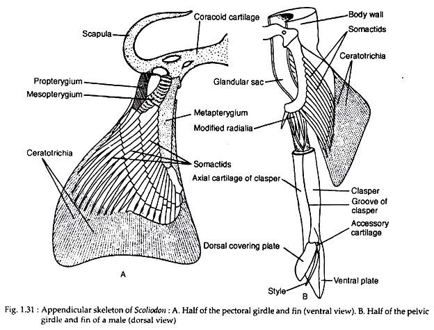
The pectoral girdle is situated posterior to the last branchial arch and consists of two semicircular cartilages united with one another along the mid-ventral line. The dorsal portion of each half is composed of a thick and rod-like scapula and the ventral portion is made up of a thin and flattened corcacoid (Fig. 1.31 A).
At the junction of the coracoid and the scapula there are three facets for the articulation of the three basal cartilages of the pectoral fin called the propterygium, mesopterygium and metapterygium, The basal cartilages bear many radial cartilages (radials) supporting the pectoral fin.
Pelvic girdle and fin:
The pelvic girdle comprises of a flattened cartilaginous rod situated along the transverse plane in front of the cloaca. A curved basal cartilage called basipterygium supports the radials of the pelvic fin (Fig. 1.31B). The basipterygium is attached anteriorly to the pelvic girdle. The radials at the distal ends contain small cartilages bearing the ceratotrichia.
In males each clasper is a tubular cartilage and grooved dorsally. Distally the groove terminates into a sharp style which is enclosed by two sheathing plates. At the upper end of the style lies a small accessory cartilage.


