In this article we will discuss about:- 1. Role of Swim-Bladder 2. Development of Swim-Bladder 3. Basic Structure 4. Types 5. Modifications 6. Relationship with Lungs 7. Relationship with Auditory Apparatus.
Role of Swim-Bladder:
In most of the fishes a characteristic sac-like structure is present between the gut and the kidneys. This structure is called by various names, viz., swim-bladder, or gas-bladder or air-bladder. The connection with the oesophagus may be retained throughout life or may be lost in the adult.
The swim-bladder occupies the same position as the lungs of higher vertebrates and is regarded as homologous to the lungs. It differs from the lungs of higher forms mainly in origin and blood supply. The swim-bladder arises from the dorsal wall of the gut and gets the blood supply usually from the dorsal aorta, while the vertebrate lung originates from the ventral wall of the pharynx and receives blood from the sixth aortic arch.
The swim-bladder is present in almost all the bony fishes and functions usually as a hydrostatic organ. Starting as a very insignificant cellular extension from the gut, the swim-bladder in fishes leads the whole group through an evolutionary channel.
Development of Swim-Bladder:
ADVERTISEMENTS:
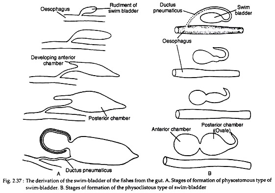
Opinions differ as regards the development of swim-bladder in fishes. In teleosts, it originates as an unpaired dorsal or dorsolateral diverticulum of the oesophagus. It starts as a small pouch budded-off from the oesophagus. The diverticulum with an opening in the oesophagus becomes subsequently divided into two halves. Of these two, the left one often atrophies except in a few primitive forms.
The right half becomes well-developed and takes a median position. In dipnoans and Polypteridae, the swim-bladder is modified into the ‘lungs’ and originates as the down-growths from the floor of the pharynx. These outgrowths have been rotated around the right side of the alimentary canal to occupy the dorsal position. As a consequence of shifting of the position, the original right positioned ‘lung’ becomes the left one.
Basic Structure of Swim-Bladder:
The swim-bladder in fishes varies greatly in structure, size and shape:
1. It is essentially a trough sac-like structure with an overlying capillary network.
ADVERTISEMENTS:
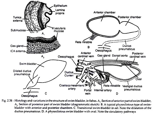
2. Beneath the capillary system the wall of the anterior part of swim-bladder consists of the following layers outside to inside (Fig. 2.38A1).
(a) Tunica externa made up of dense collagenous fibrous material.
(b) Sub-mucosa, consisting of loose connective tissue.
(c) Muscularis mucosa, consisting of a thick layer of smooth muscle fibres.
ADVERTISEMENTS:
(d) Lamina propria, formed of thin-layer of connective tissue and
(e) Innermost layer of epithelial cells.
3. In the posterior chamber of swim bladder, outside the layer of muscularis mucosa there is a glandular layer. This layer is richly supplied with blood capillaries from rete mirabile (Fig. 2.38A2).
4. The swim bladder opens into the oesophagus by a ductus pneumaticus, which is short and wide in lower teleosts (Chondrostie and Holostei), while in others it is longer and narrower. The gas secreted by the swim- bladder is mostly oxygen. Nitrogen and little quantity of carbon dioxide are also present.
Types of Swim-Bladder:
Depending on the presence of the duct (ductus pneumaticus) between the swim-bladder and the oesophagus, the swim-bladder in fishes can be divided into two broad categories: Physostomus and Physoclistous types. Depending on the condition of the swim-bladder, the teleosts are classified by older taxonomists into two groups Physostomi and Physoclisti. A transitional condition is observed in eels.
A. Physostomous Condition:
The swim-bladder develops from the oesophagus. When the ductus pneumaticus is present between the swim-bladder and the oesophagus, the swim-bladder is called physostomous type (Fig. 2.37A). A vessel emerging from the coeliacomesenteric artery supplies the swim-bladder and the blood from it is conveyed to the heart through a vein joining the hepatic portal vein. This condition is observed in bony fishes, the dipnoans and soft-rayed teleosts.
B. Physoclistous condition:
ADVERTISEMENTS:
In this condition the ductus pneumaticus is either closed or atrophied (Fig. 2.37B). This type of swim- bladder is observed in spiny-rayed fishes. In this type of swim-bladder, there lies an anteroventral secretory gas gland (containing retia mirabilia) and a posterodorsal gas-absorbing region called the ovale.
The oval develops out of the degenerating ductus pneumaticus. The rete mirabilis of the gas gland, the oval and the walls of the bladder are supplied by the coeliacomesenteric artery and also by arteries from the dorsal aorta. But the blood from the different parts of the swim- bladder is returned by two routes.
The blood from the gas gland is returned to the heart by the hepatic portal vein, while from the rest of the bladder by the posterior cardinal veins. The bladder, specially the gas gland, gets the lateral branches from the vagus, while the oval is innervated by sympathetic nerves. This condition is present in some dipnoans.
C. Transitional condition:
In Eel (Anguilla), a transitional condition between the physostomous and physoclistous type is present. The swim-bladder retains the ductus pneumaticus which becomes enlarged to form a separate chamber containing the oval (Fig. 2.38C).
The gas glands are also present. The swim-bladder is supplied with the blood through a branch from the coeliacomesenteric artery while the blood is returned to the heart by a vessel joining the post-cardinal vein. The condition represents an intermediate stage when a physostomous condition is on the verge of transformation into the physoclistous state.
Modifications in Swim-Bladder:
In fishes a great diversity in size, shape and function of the swim-bladder is observed. In elasmobranchs, bottom dwelling and deep-sea teleosts the swim-bladder is absent in the adult but a transitory rudimetit during development may be present.
In flat fishes (Pleuronectidae) swim-bladder is present in the early life when the animals maintain a vertical position. As they tip over one side and assume the lazy adulthood, the swim-bladder becomes atrophied.
In elasmobranchs, the swim-bladder is represented by the transitory rudiment in the embryonic stages. One to six oesophageal pits in Pristiurus, Torpedo and Trygon are recorded. These pits are located posterior to the fifth pouch.
In sharks the swim-bladder is absent in adults, but a hint of a rudimentary swim- bladder is observed during embryonic development. But almost all the teleosts possess the swim-bladder and extreme modifications of the same are encountered because of adaptation to the different modes of living.
A. Modifications of physostomous condition:
The typical physostomous pattern becomes modified in different fishes and the basic trends are:
(1) The formation of paired sacs, and
(2) The gradual acquisition of two chambers — an anterior and a posterior.
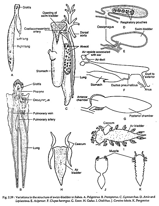
The swim-bladder varies extensively in size and shape. In some forms it gives off many branched diverticula. The swim-bladder in Polypterus (Fig. 2.39A) represents the primitive condition.
It is a bilobed sac with two unequally developed lobes. The left lobe is shorter and the right lobe is longer. The bilobed sac opens on the floor of the pharynx through a slit-like glottis. The glottis is provided with muscular sphincter. The internal lining of the bladder is smooth and partly ciliated.
The lack of alveolar sacculations and the presence of muscular walls are the two noted features in the swim-bladder of Polypterus. The walls of the bladder are highly vascular and are lined by two layers of striated muscle fibres. The bladder is supplied by a pair of pulmonary arteries arising from the last pair of epibranchial arteries and the corresponding veins enter into the hepatic vein below the sinus venosus.
In the dipnoans, the swim-bladder is called the lung and the inner walls are produced into numerous alveoli. The swim-bladder resembles the tetrapod lungs both structurally as well as functionally. In Neoceratodus it is single-lobed, while in Protopterus and Tepidostiren it is bilobed (Fig. 2.39B). Other details regarding the structural construction, blood and nerve supplies have already been dealt in the biology of the lung-fishes.
In Acipenser (Fig. 2.39E), the swim-bladder is short and oval in shape. The ductus pneumaticus enters the bladder ventrally and it opens into the gut posterior to the pharynx. The glottis is lacking and the opening into the oesophagus is closed by the simple constriction of the ductus pneumaticus.
The walls of the bladder are fibrous and thick but the inner walls are smooth. In Acipenser, both the left and right lobes develop from the dorsal side of the oesophagus in the embryonic stage, but the left one becomes completely obliterated and right one gives rise to the adult swim-bladder.
In Amia and Lepisosteus (Fig. 2.39D), the swim-bladder is an unpaired sac extending nearly the entire length of the body cavity. In both the cases rudiment of the left lobe appears during development but persists only for a short time. The ductus pneumaticus opens into the oesophagus posterior to the pharynx through a dorsal slit-like glottis. The walls are highly vascular and exhibit sacculations resembling the pulmonary alveoli of Protopterus.
The sacculations or the respiratory pouches are arranged in two lateral rows. As regards the development of sacculations, the swim-bladder of Lepisosteus is more advanced than that of Amia. There are some more minor differences regarding the supply of blood. The swim-bladder in Amia gets arterial blood from the pulmonary arteries, while that of Lepisosteus gets arterial branches from the dorsal aorta.
The blood from the bladder is returned by the left ductus Cuvieri in Amia and by the right post-cardinal in Lepisosteus. Gymnarchus (Fig. 2.39C) presents an intermediate stage where the efferent branchial arteries from the third and fourth gill-arches join to form a common root for the emergence of the pulmonary and coeliacomesenteric arteries. Amongst the dipnoans, the swim-bladder of Neocertatodus resembles that of Lepisosteus. The walls are sacculated and act as the ‘lung’.
In Clupea harengus (Fig. 2.39F) the ductus pneumaticus opens into the fundus of the stomach and there is a second duct from the posterior part of the swim-bladder opening to the exterior near the anus. Similar posterior opening is present in Pellona, Caranx, Sardinella.
In many fishes, the anterior prolongations of the swim-bladder come into close contact with the wall of the space containing the internal ear. In Clupea, the narrow anterior end of the swim-bladder enters into a canal in the basioccipital of the skull and divides into two slender branches.
The anterior end of each branch dilates to form a round-swelling and lies in close contact with the internal ear. A more or less similar condition is observed in Tenulosa ilisha.
In many fishes, finger-like diverticula develop from the swim-bladder. In Gadus, a pair of diverticula originating from the anterior part of the bladder project into the head region (Fig. 2.39H). In Otolithus, each anterolateral end of the swim-bladder gives rise to an outgrowth which sends one anterior and a posterior horn (Fig. 2.391). In Corvina lobata, many such branched diverticula develop from the lateral walls of the swim-bladder (Fig. 2.39J).
Usually in most cases, the swim-bladder is divided transversely into an anterior and a posterior chamber as seen in cyprinoids, Essox, Catostomus, Pangassius (Fig. 2.39K), Corvina, etc. But the longitudinal division of the swim- bladder is rare.
In Arius the swim-bladder is splitted longitudinally. In Notopterus, a longitudinal septum divides the swim-bladder into two lateral chambers. Due to the presence of septum or septa, the internal cavity of the swim-bladder is either completely or partially divided.
B. Modifications of physoclistous condition:
The swim-bladder in all teleosts begins as a physostomous type but in an adult condition the ductus pneumaticus gets degenerated to become a physoclistous type. A typical physoclistous swim-bladder consist of a closed sac having two compartments—an anterior and a posterior. These two compartments are inter-communicated through an aperture called ductus communicans.
The opening and closure of this aperture is regulated by circular and radiating muscles which act as the sphincter. The anterior chamber is formed by the enlargement and forward growth of the budding swim-bladder, while the posterior chamber develops as an enlargement of the ductus pneumaticus. This typical structural plan is modified in certain forms.
The posterior chamber with retina mirabilia becomes flattened almost to the point of obliteration and is designated ‘ovale’ as seen in the families like Myctophidae, Percidae, Mugilidae. The oval is a thin-walled highly muscular area specialised for the reabsorption of gases (see Fig. 2.38D). The opening of the oval is guarded by circular and longitudinal muscles. This device is of great significance for the fishes undergoing rapid vertical movements.
Histological Modifications:
The morphological modifications of the swim-bladder are accompanied by histological modifications in different fishes, the swim-bladder acts as a hydrostatic organ. It helps fishes to sink or ascend to various depths by altering the gas content in the bladder. In fishes having open ductus pneumaticus, the volume of gas content in the bladder can be changed by swallowing or removing air from the bladder.
But in some physostomous and all physoclistous fishes, this process of gas transference is done directly from the blood stream. Inside the bladder there is an oxygen-producing device and an oxygen-absorbing device. The swim-bladder is a vascular structure but the degree of vascularization varies in different teleosts.
In some species of the families Clupeidae and Salmonidae the capillaries are uniformly present all over the swim-bladder, but in most cases these highly vascular interlacing and tightly packed capillaries form a mass called rete mirabilis (Fig. 2.38D). The anterior chamber of swim-bladder shows the tendency to become differentiated into oxygen-producing area called red body.
Oxygen is produced by the reduction of the oxyhaemoglobin in the erythrocytes when brought into close contact with the secreting epithelial cells of the gas gland. The red body consists of an internal oxygen-secreting cells (gas gland) and supplied by the blood vessels from the reta mirabilia (singular, rete mirabilis).
It forms a complicated structure where the arterial and venous capillaries communicate only after reaching the gas gland. The most primitive condition is observed in Pickerel where the gland is covered by thick glandular epithelium which is thrown into a number of folds. In eels and some other fishes, the red bodies are non- glandular in nature but serve the same physiological function.
The red gland is supplied with blood from the coeliac artery and is returned to the portal vein. The activity of the red gland is controlled by the vagus nerve. In the fishes with functional ductus pneumaticus the gas gland is absent but in eels this function is taken up by the red gland.
In the physoclistous fishes, the anterior region is modified for gas production and the posterior region or chamber is specialised for the absorption of gas into the blood. The posterior chamber becomes excessively thin-walled to facilitate gas diffusion. Beneath the walls, the gas is absorbed directly into the blood. The formation of the ovale in some fishes is a special development for the absorption of gas.
The wall of the ovale is very thin and highly vascular. Through this epithelial lining oxygen can easily pass to the network of vessels. This gas-absorbing region receives blood supply from the dorsal aorta and the blood is returned to the post-cardinal vein. The activities are governed by the sympathetic nerves.
The histological differentiation for the gas production and gas absorption is a very significant achievement in fishes. The gas produced by the red body is mostly oxygen and this oxygen is readily absorbed or diffused from the swim-bladder directly into the capillaries. The oval is modified for gas absorption in many fishes.
By the alternate processes of gas production and gas absorption, the internal pressure and volume of the gas content inside the swim-bladder can be increased or decreased. The red body is usually confined to the anterior chamber, but in fishes where the anterior chamber becomes secondarily associated with the auditory function, the gas gland may be confined to the posterior chamber.
Functions of Swim-Bladder:
The swim-bladder in fishes performs a variety of functions.
Hydrostatic organ:
It is primarily a hydrostatic organ and helps to keep the weight of the body equal to the volume of the water the fish displaces. It also serves to equilibrate the body in relation to the surrounding medium by increasing or decreasing the volume of gas content.
In the physostomous fishes the expulsion of the gas from the swim-bladder occurs through the ductus pneumaticus, but in the physoclistous fishes — where the ductus pneumaticus is absent —the superfluous gas is removed by diffusion.
Adjustable float:
The swim-bladder also acts as an adjustable float to enable the fishes to swim at any depth with the least effort. When a fish likes to sink, the specific gravity of the body is increased. When it ascends the swim-bladder is distended and the specific gravity is diminished. By such adjustment, a fish can maintain an equilibrium at any level.
Maintain proper centre of gravity:
The swim-bladder helps to maintain the proper centre of gravity by shifting the contained gas from one part of it to the other and thus facilitates in exhibiting a variety of movement.
Respiration:
The respiratory function of the swim-bladder is quite significant. In many fishes living in water in which oxygen content is considerably low, the oxygen produced in the bladder may serve as a source of oxygen. In few fishes, specially in the dipnoans, the swim-bladder becomes modified into the ‘lung’. The ‘lung’ is capable of taking atmospheric air.
Resonator:
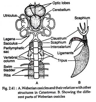
The swim-bladder is regarded to act as a resonator. It intensifies the vibrations of sound and transmits these to the ear through the Weberian ossicles.
Production of sound:
The swim-bladder helps in the production of sound. Many fishes, Doras, Platystoma, Malapterurus, Trigla can produce grunting or hissing or drumming sound. The circulation of the contained air inside the swim-bladder causes the vibration of the incomplete septa. The sound is produced as the consequence of vibration of the incomplete septa present on the inner wall of the swim- bladder.
The vibrations are caused by the movement of the contained air of the swim- bladder. Sound may also be produced by the compression of the extrinsic and intrinsic musculatures of the swim-bladder. Polypterus, Protopterus and Lepidosiren can produce sound by compression and forceful expulsion of the contained gas in the swim-bladder. In Cynoscion male, the musculus sonorificus probably helps in compression.
Relationship between Swim-Bladder and Lung:
The swim-bladder in fishes and the lungs of tetrapods are closely similar in structure and development. Both the structures are intimately associated with the gut. The lungs of higher vertebrates arise exactly the same way as that of the swim-bladder in fishes. Both develop as outgrowths from the gullet and the glottis occupies exactly the same position.
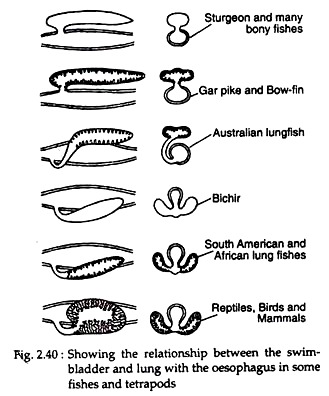
But the exact nature of such similarity becomes difficult to estimate because of the lack of connecting fossil records. The evidences advanced by the study of embryology and comparative anatomy make the data more confusing. Figure 2.40 shows the relationship of swim-bladder of a few fishes and lungs in higher vertebrates with the gullet.
The swim- bladder in fishes shows a gradual sequence of evolution from the simple to complex and there is general tendency to become lung-like in structure and function. In dipnoans the swim-bladder is actually a true air-breathing organ. In the primitive stage as shown by the sturgeon and many other fishes, the swim-bladder is a simple sac-like structure filled with a mixture of gases and sub-serves hydrostatic function.
But in Amia, Lepisosteus and dipnoans the inner wall of the swim-bladder is richly vascularized and the surface area is increased by the development of pulmonary alveoli. In most cases, the swim-bladder is a single structure, but in Polypterusy. Amia, Lepisosteus, Protopterus and Lepidosiren the swim-bladder is paired (unequally developed in Polypterus).
The right portion is long and the left one is of much shorter structure and opens by a single slit-like aperture (glottis) into the gullet. The arrangement of blood vessels to this particular organ, specially in the dipnoans, is very similar to that of the amphibians.
Relation between Swim-Bladder and Auditory Apparatus:
The swim-bladder, in many fishes, becomes intimately connected with the internal ear. This connection possibly enables the fishes to transmit change of pressure to the perilymph. The association between swim-bladder and auditory apparatus exhibits a gradual trend of evolution from the simple to complex.
In Gudus, Megalopa and others, this connection is very simple. A finger-like diverticulum, after originating from the anterior end of the swim-bladder, joins the membrane covering an opening of the auditory capsule. In Clupeidae, the situation is more advanced and a pair of such tubular outgrowths enters inside the auditory capsule.
In Clupeidae, the situation is more advanced and a pair of such tubular outgrowths enters the auditory capsule. Each branch again divides into two, and terminates to a swollen vesicle. These vesicles are enclosed inside the prootic and pterotic bones. The vesicles touch a process coming from the membranous labyrinth.
But in carps and Siluroids (order Ostariophysi), this connection is of more advanced nature. The connection is made by a chain of ossicles called the Weberian ossicles (named after the discoverer Weber). These ossicles are not homologous to the ear ossicles of other vertebrates, but are formed by the specialisation of few anterior vertebral segments.
From the above facts, it is apparent that the swim-bladder and the lungs of the tetrapods are strikingly similar in origin, structure and function. Existence of such close parallelism led many workers to think that the swim-bladder of fishes is the forerunner of the tetrapod lung in the phylogenetic history of the vertebrates.
But such an idea is not supported by the geological evidence. The appearance of lung is geologically more primitive than the swim-bladder of fishes. This occurrence rejects the plea of the derivation of the lung directly from the swim- bladder, but their monophylectic origin is a very popular notion.
It is regarded that the placoderms constitute the most primitive gnathostomes and they possess swim-bladder. The lungs of tetrapods and the swim-bladder of modern fishes have evolved from the placoderm source. The idea appears very simple to interprete, but the actual records of evolution are lacking.
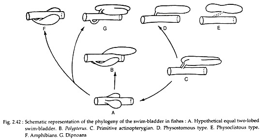
Diverse opinions exist on this particular issue. The most recent trend is to assume that the swim-bladder originated from a paired pouches which became fused into one in the course of evolution (Fig. 2.42). This condition is observed in fishes where the swim-bladder is simply hydrostatic in function.
The swim-bladder of Polypterus, Protopterus and Lepidosiren presents fairly the primitive condition because in all of them the swim-bladder is paired and transformed into the ‘lungs’. Now the question arises regarding the relationship between hydrostatic and respiratory types of the swim-bladder.
Fishes having respiratory swim-bladder (Polypterus, Protopterus and Lepidosiren) cannot be regarded as the progenitors of the teleosts possessing the swim-bladder of hydrostatic type. So it is logical to think that fishes having the hydrostatic type of swim-bladder evolved independently. Nothing more can be enlightened on this particular aspect, as our knowledge about the swim-bladder of the crossopterygians is insufficient.
