The digestive system of frog mainly includes the alimentary canal and the digestive glands. In the alimentary canal the processes of mastication, digestion and absorption take place, while the digestive glands secrete certain enzymes which bring about the digestion of the ingested food.
Alimentary Canal:
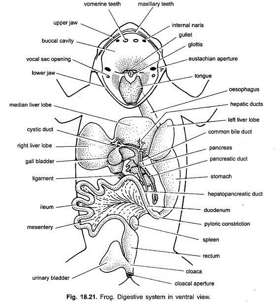
The alimentary canal of the frog is essentially a coiled tube of varying diameter that extends from mouth to cloacal aperture. It consists of the mouth, buccal cavity, pharynx, oesophagus, stomach, intestine and cloaca.
1. Mouth:
The alimentary canal starts with an aperture which is known as mouth opening. Mouth is a very wide gape bounded by upper and lower jaws covered by immovable lips.
ADVERTISEMENTS:
2. Buccal Cavity:
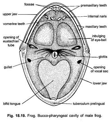
Mouth leads into a wide and broad oral or buccal cavity which lies in between the two jaws-the upper and the lower jaw. The upper jaw is fixed or immovable, while the lower jaw is hinged and can be freely moved up and down in the vertical plane. The buccal cavity is lined by ciliated, columnar epithelium which contains mucus secreting glands. Mucus lubricates the food. In frog, salivary glands are absent.
(i) Teeth:
ADVERTISEMENTS:
The upper jaw bears a row of closely set, small, uniform and hook-like pointed teeth over the premaxillae and maxillae, but the lower jaw lacks the teeth.
Besides these, there are two more patches of teeth found one on either side of the median line of the roof of the buccal cavity, they are called vomerine teeth as they are born on the vomer. These teeth are not used for chewing but prevent the escape of the captured prey.
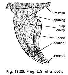
The teeth are homodont, i.e., similar in shape. They are curved backwards and attached to the bones (acrodont) instead of being set in sockets. Each tooth is conical in its shape and consists of two parts-the base and the crown. The base is attached to the jaw bone and is made of a bone-like substance.
The crown is the free end of tooth and is formed of dentine traversed by numerous fine branching canals or canaliculi leading from the interior of the tooth. The tip of the crown is coated with a very hard, resistant, shining white layer of enamel substance. In the interior of each tooth there is a cavity which is called pulp cavity.
ADVERTISEMENTS:
It is filled with a highly vascular and nutritive tissue or pulp which also contains odontoblast cells which produce new material for the growth of teeth. During lifetime the old and worn out teeth are being continuously replaced by new ones which are produced below the old ones which are cast out or lost. As the teeth are replaced several times during the lifetime of the frog, so they are called polyphyodont.
(ii) Internal Nostrils:
The buccal cavity in its roof near the vomerine teeth has two openings, the internal or posterior nares connecting with the nasal cavities through which the respiratory gases pass to and from the buccal cavity during respiration.
(iii) Tongue:
The large, thick, fleshy and protrusible tongue attached in front to the inner border of lower jaw, and free and notched behind lies on the floor of the buccal cavity. Its upper surface bears taste buds in the form of small papillae and mucous glands of which the secretions make the tongue sticky. Neither taste buds nor mucous glands produce any digestive enzymes.
The tongue can be thrown out and retracted suddenly to capture and engulf insects. According to Hartog, the throwing out of the tongue is brought about by the sudden flowing of squeezed lymph from the lymph sac to another due to muscular contraction but the swallowing is accomplished simply by raising the floor of the buccal cavity in which a flat hyoid cartilage is embedded.
(iv) Bulgings of Orbits:
The roof of buccal cavity behind the vomerine teeth has two large, oval pale areas, the bulgings of eyeballs. During swallowing of food, eyes are depressed into the buccal cavity which pushes the food into the pharynx.
ADVERTISEMENTS:
3. Pharynx:
The buccal cavity narrows behind as the pharynx which in turn opens into the oesophagus through gullet. The buccal cavity and pharynx are sometimes called the buccopharyngeal cavity. In the roof of the pharynx on either lateral side is present a wide eustachean opening which communicates with the middle ear.
The glottis which is a median slit in the pharynx behind the tongue guards the entrance to the lungs. It is always opened during breathing but closes while food is being swallowed. In male frog two openings of vocal sacs are also formed in the angle of the lower jaw on the floor of the pharynx. These act as resonators at the time of croaking.
4. Oesophagus:
The gullet leads into a short, broad and muscular part of the alimentary canal called oesophagus. This part of the alimentary canal is very short due to the absence of neck but highly distensible as its inner lining is thrown into a large number of longitudinal folds which allow the sufficient expansion of the oesophagus during the passage of the ingested food through it to the stomach. It opens into the stomach in such a way that no demarcation line is formed between the oesophagus and stomach.
5. Stomach:
It is the most important part of the alimentary canal where the digestion of ingested food takes place with the help of certain digestive enzymes secreted by the digestive glands situated in its wall. It is in the form of wide and curved tube and lies between the oesophagus and intestine.
It is suspended by a mesentery, the mesogaster, into the left side of the body cavity or the coelom. It is distinguished into two parts – the large wider anterior cardiac stomach and the posterior, short narrow pyloric stomach.
The internal lining of the stomach has numerous longitudinal folds which may allow the expansion of the stomach whenever needed. Its mucous epithelium possesses multicellular gastric glands which secrete pepsinogen enzyme, and unicellular oxyntic glands which secrete hydrochloric acid.
The pyloric end of stomach is constricted and its opening into small intestine is guarded by sphincter (a circular ring-like muscle). It regulates the passage of food from the stomach into the intestine.
6. Intestine:
The stomach leads into the long, tubular and coiled intestine. It is also attached to the dorsal body wall by mesentery.
It consists of two parts:
a. The small and
b. Large intestine.
a. Small Intestine:
The small intestine lies in several loops supported by a fan-like membrane, the mesentery. The anterior region of the small intestine which curves upwards to form a U with the stomach is the duodenum, the rest part of it continues as the coiled ileum. In the duodenum opens a common hepato pancreatic duct from liver and pancreas bringing the bile and pancreatic juice. The internal mucous lining is thrown into low transverse folds.
b. Large Intestine:
The ileum is a long, narrow, very much coild tube and its lower end leads into rectum (large intestine). The internal mucous lining of ileum forms a number of longitudinal folds. True villi, glands and crypts of higher vertebrates are absent. Digestion and absorption of food takes place in this part. Its lower end opens into cloaca by spinctered anus. Its mucosal lining forms low longitudinal folds.
7. Cloaca:
It is a small sac-like structure and receives the openings of anus and urinogenital apertures. Cloaca opens towards the exterior through vent or cloacal spening situated at the posterior end of the body.
Digestive Glands:
Two large digestive glands, in addition to the glands located in the stomach and small intestine, are also found in the frog. These glands are the liver and the pancreas.
(1) Liver:
It is the largest reddish brown gland found in the anterior region of the body cavity close to the heart and lungs. It consists of two lobes-the right and the left. These two lobes remain connected with each other by a narrow bridge of liver tissue.
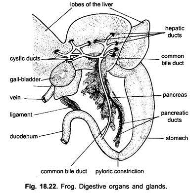
The left lobe is again subdivided into two lobes. Between the right and left lobes, a thin-walled, round, large, greenish sac, the gall bladder, is found. It serves as a reservoir for the bile constantly secreted by the liver cells.
The bile passes into the gall bladder through cystic ducts as well as directly into the bile duct by way of minute hepatic ducts. The hepatic and crystic ducts join to form a common bile duct which runs through the pancreas and opens into the duodenum.
The common bile duct is also known as hepatopancreatic duct as it receives the fine ducts of the pancreas on its way to the duodenum. Bile lacks digestive juices. Bile only emulsifies fats, thus, liver is not a true digestive gland.
Histology of Liver:
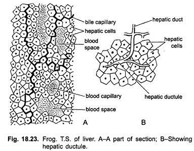
The liver is composed of numerous lobules or tubules which not only branch but also anastomose with another to form a complex network due to which it is sometimes called as a reticular gland. These lobules are separated from each other by the presence of connective tissue containing bile capillaries, hepatic ducts, blood sinuses and blood capillaries.
Each lobule is made up of numerous polyhedral, glandular hepatic cells which contain nuclei and cytoplasm along with protein granules, droplets of fats, glycogen and often black or dark brown pigment granules. The hepatic cells are arranged in columns between the bile capillaries which invite to form larger hepatic ducts. Hepatic ducts ultimately open into cystic duct directly attached with the gall bladder. The hepatic ducts of different lobes and cystic duct unite to form the bile duct.
The liver receives blood from the hepatic artery and the hepatic portal vein. These blood vessels enter the liver and provide the required material for the formation of bile. The bile constantly secreted by the liver cells exudes into capillaries from which it passes either into the gall bladder via cystic ducts for the temporary storage or the intestine through the bile duct.
Functions of Liver:
The liver performs the following important functions:
1. The liver secretes a watery, alkaline bile which contains bile salts, bile pigments, cholesterol, lecithin and water. Bile salts are bicarbonate, glycocholate and taurocholate of sodium. Sodium bicarbonate reduces the acidity of food in the intestine, while the other two bile salts activate the pancreatic lipase and lower the surface tension of fats so that they can be emulsified.
2. It has no digestive enzymes but it adds water to the food and aids in the digestion of fats by emulsifying them.
3. It stores excess sugar in the form of glycogen, which is formed by the change of glucose (glycogenesis). It is stored as a reserve food but it can be changed into glucose (glycogenolysis) when its concentration falls in the blood.
4. It maintains the protein concentration in blood. The excess amino acids instead of being stored are converted into ammonia by liver, which combines with carbon dioxide and is changed into urea and other nitrogenous wastes by the action of enzymes (deamination) which are finally eliminated as urine by the kidneys.
5. It also removes some other excretory products which are discharged with bile into the intestine and expelled with the faeces.
6. In an embryo the liver forms red blood corpuscles, but in an adult it destroys old and worn out erythrocytes by its Kupffer cells.
7. It stores copper and iron and forms vitamin A.
8. It produces fibrinogen and prothrombin which are essential for the clotting of blood. It also produces heparin which prevents clotting of blood in blood vessels.
9. It kills bacteria and also eliminates foreign substances from the blood.
10. Prussic acid is formed in the body as a by-product and it is harmful. Liver converts it into harmless potassium sulphocyanide.
(2) Pancreas:
It is an irregular, branched, flattened and pale coloured gland of exocrine and endocrine function, which is held by mesentery between the stomach and duodenum. It is traversed by the common bile duct into which the pancreatic ducts also open which is now called as hepatopancreatic duct.
Exocrine Part:
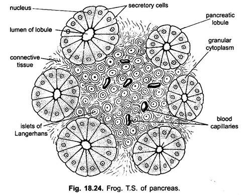
It is divided into many lobes and lobules held together by connective tissue in which are present pancreatic ducts, blood vessels, lymph vessels and nerves. The lobules have numerous branching tubules or acini or alveoli. Each alveolus is formed of pyramidal glandular pancreatic cells around a central cavity.
These alveoli unite with each other through their ductules, which in turn unite to form the larger ducts and then finally form the pancreatic ducts. The pancreatic ducts open into the bile duct when it traverses the pancreas. The pancreatic cells have large nuclei and non-granular cytoplasm. The pancreas produces pancreatic juice which contains various enzymes which digest the proteins, carbohydrates and fats of the ingested food.
Endocrine Part:
Amongst the acini in the connective tissue are present compact groups of cells which are known as pancreatic islets or islets of Langerhans. These cells are somewhat spherical and arranged in compact groups and take light stain. There are three kinds of cells in an islet separated by capillaries.
(i) Alpha Cells:
These cells have achromatic nuclei and large acidophilic granules. These cells secrete glycogen hormone, which increases the sugar concentration in blood. Its deficiency results in hypoglycemia.
(ii) Beta Cells:
They possess small, rounded, deeply stained nuclei and orange-brown granules. They secrete insulin hormone.
(iii) D Cells:
These cells possess vesicular nuclei and basophilic granules.
Insulin is essential for metabolism of carbohydrates, regulating the storage of glycogen in liver and muscles, concentration of sugar in the blood and for increasing the ability of tissues to oxidise glucose as a source of energy. Deficiency of insulin causes a disease called diabetes (hyperglycemia).
Histology of the Alimentary Canal:
Histologically, the wall of alimentary canal of frog and other vertebrates is made up of four distinct concentric layers.
These are present in the following order from within-outwards:
(1) The mucosa;
(2) The submucosa;
(3) The muscularis; and
(4) the visceral peritoneum or serosa.
1. Mucosa:
It is the innermost layer or mucous membrane. It remains folded forming various pits and different types of glands. It is concerned with secretion and absorption of digested food, etc.
It is further composed of the following layers:
(i) Epithelium:
It is the innermost layer formed of simple columnar epithelium (glandular and ciliated) based on thin basement membrane.
(ii) Lamina Propria:
It is a thin connective tissue layer having blood capillaries, lymph and nerves.
(iii) Muscularis Mucosae:
It is a narrow smooth muscle layer containing inner circular and outer longitudinal muscles.
2. Submucosa:
It is a thin protective layer composed of coarse connective tissue, elastic fibres, fat, blood and lymph vessels and nerve cells. It contains glands in mammal. It also possesses Meissner plexus formed of nerve cells and fibres.
3. Muscularis:
It is composed of outer longitudinal and inner circular smooth muscle fibres which are in spirals. Between these two layers of muscles is a thin layer of connective tissue having a network of nerve cells and nerve fibres of the autonomic ganglionated myocentric Auerbach plexus.
4. Visceral Peritoneum or Serosa:
It is the outermost thin layer which lacks in the oesophagus. It is composed of very much thin connective tissue layer and an outer layer of flattened cells. It is continuous with the peritoneum lining of the body cavity called mesentery.
Certain variations are found in different parts of the alimentary canal because the above said layers, particularly the mucosa, become greatly modified in different parts of the alimentary canal to enable that part to carry on certain specific functions.
a. Oesophagus:
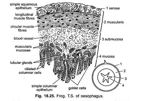
Histologically, the oesophagus more or less has the same structure, but it differs from the rest of the alimentary canal in the following facts:
(i) It has no visceral peritoneum because it lies outside the coelom.
(ii) It has both voluntary and involuntary muscle fibres in its wall and voluntary or striated muscle fibres in the upper portion.
(iii) Its mucous membrane lining is made of stratified squamous epithelial cells instead of columnar cells.
It has some goblet cells which produce mucus making the food material slippery. Mucous epithelium is invaginated into submucosa forming tubular branched glands secreting an enzyme pepsin for the digestion of proteins.
b. Stomach:
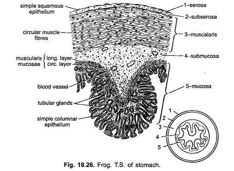
The wall of the stomach (Fig. 18.26) is thick and made of the same typical parts of the alimentary canal, i.e., mucosa, submucosa, muscularis and serosa, but it has two peculiarities:
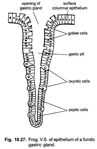
(i) Stomach wall is thick and longitudinally folded internally. Folds disappear when stomach is distended. The mucous epithelium is formed of simple columnar mucous secreting gland cells. These glands are embedded in the connective tissue of lamina propria. Glands represent invaginations of the mucous epithelium. They are elongated tubular structures set very closely together, and frequently more or less branched, called gastric glands.
(ii) These glands differ in their structure at different regions of the stomach. The gastric glands of the cardiac region of the stomach are called cardiac glands, while of the fundus and pyloric region are called as fundic and pyloric respectively.
The cardiac glands are very long with deep set mouth but the fundic and pyloric glands comparatively are less deep and smaller. The cardiac and pyloric glands secrete only mucus from the surface cells. Fundic glands (or cardiac glands in some) have three kinds of cells, mucus neck cells produce mucus, oxyntic cells produce hydrochloric acid, they may be present in the cardiac region also, zymogen cells or peptic cells produce pepsin. Pepsin is produced in the form of inactive propepsin or pepsinogen which is soon converted into active pepsin by the hydrochloric acid.
(iii) Villi are absent.
c. Small Intestine or Duodenum:
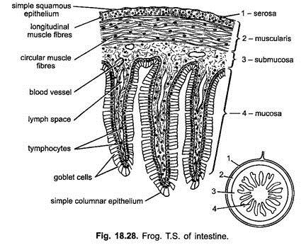
It has all the four usual coats of the alimentary canal, but its mucosa is quite thick which forms irregular, branched transverse folds which increase the absorptive surface of the alimentary canal. It is formed of columnar epithelial cells having mucous secreting gland cells. Mucosa of small intestine is thrown into numerous folds, but there are no true villi nor definite glands nor crypts of higher vertebrates. Muscularis mucosae is thin. Remaining layers are of typical type.
Ileum:
It also contains usual four coats. Mucosa is thrown into several folds of various sizes and these are produced into the intestinal lumen decreasing its diameter. Mucosa has tall columnar epithelial cells in a single row within which goblet and absorptive cells are scattered. The muscle layers are less developed. Muscularis mucosa is thin and comprises only one layer of muscle fibres. True villi, crypts are lacking.
d. Large Intestine or Rectum:
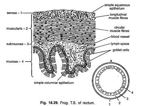
It is also composed of the same four coats of the alimentary canal. The muscularis mucosae is less developed, muscular coat is thick and both contain voluntary muscle fibres. The mucosa is thick and forms longitudinal folds and comprises numerous tubular goblet cells, secreting mucus. Mucosa epithelium is stratified near anus.
Physiology of Digestion:
1. Food:
The frog is carnivorous, feeds chiefly on earthworms, spiders, snails, fishes, smaller frogs and other small insects which it captures and swallows whole directly into the stomach with the help of protractible tongue.
2. Ingestion of Food:
Its mode of catching of its prey is remarkable. When feeding, the frog sits at a suitable place frequented by insects. When any prey comes near to it, it opens its mouth and suddenly flicks out its sticky tongue and strikes the prey. As soon as the prey comes in contact with the tongue it immediately adheres to it. Now the tongue is withdrawn into the buccal cavity.
Once the prey is caught into the buccal cavity it would not be allowed to escape due to the presence of the hook-like inwardly directed maxilliary and vomerine teeth. From the buccal cavity it is pushed into oesophagus by the contraction of the pharyngeal wall and thence by peristalsis caused by the contraction and dilation of muscular wall of oesophagus, into the stomach.
3. Digestion of Food:
During feeding whatever the food is ingested by the animal that contains complex organic substances which are not of immediate use as they are insoluble and cannot diffuse through the mucous membrane lining of the alimentary canal, therefore, they must be subjected to the physical and chemical changes of the digestion so that they may be transformed into soluble forms for the immediate use of the body.
The physical changes are brought about by the peristaltic movements of the alimentary canal, while the chemical changes are brought about by the organic catalysts called enzymes which only hasten the chemical reactions without being changed themselves.
They are always specific in their property and are complex proteins which are produced by the exocrine glands and they always act at the optimum body temperature. They can also reverse the reaction, i.e., the substances they had changed can be reformed. They are of different types subject to the type of food on which they act. Thus, proteins are digested by proteolytic enzymes, carbohydrates by diastatic (amylolytic) enzymes and fats by lipolytic enzymes.
(i) Buccal Digestion:
In frog the captured prey is neither subjected to any physical change (mastication) nor any chemical action in the buccal cavity as the buccal epithelium does not have any digestive gland.
From the buccal cavity the prey is directly pushed into oesophagus where it undergoes physical changes due to constant peristaltic movement of its wall. Besides mucus, the glands of oesophagus also secrete an enzyme called pepsin but no digestion occurs as it does not become active till it reaches the stomach, while the mucus simply makes the active food inactive and soft and, thus, makes the passage easier.
(ii) Gastric Digestion:
Due to oesophageal contractions and relaxations the food comes down to stomach.
The stomach performs the following three main functions:
(a) Storage;
(b) Mechanical mixing; and
(c) Chemical modifications.
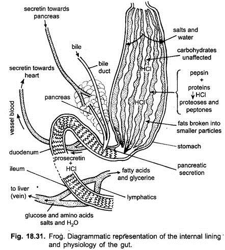
As soon as food reaches the stomach, its wall shows a series of muscular contractions, the so-called peristaltic movements which not only allow the food to go down but also break down into small pieces and mix it thoroughly with gastric juice secreted by the gastric glands situated in the internal lining of stomach. The gastric glands secrete their secretions when they are activated by gastrin hormone which is produced by the stomach wall as soon as the food comes down to stomach.
The gastric juice secreted by the gastric glands contains a large amount of water, inactive pepsinogen enzyme and free hydrochloric acid. Inactive pepsinogen changes into active pepsin on being mixed with hydrochloric acid. The acid also prevents the bacterial decomposition and dissolves the inorganic salts as well as makes the food soft. The pepsin of stomach along with pepsin of oesophagus acts on proteins of food and changes them into peptones and proteoses.
Pepsinogen + HCl → Active pepsin
Pepsin + Proteins → Peptones + Proteoses
After being remained for about 2-3 hours in the stomach the food is thoroughly churned and mixed by the muscular contraction of the stomach wall and takes the form of a thick creamy acid fluid called chyme. Muscular contractions of the stomach wall force the chyme to pass in small amount at a time through the pylorus into the duodenum.
(iii) Intestinal Digestion:
The food as it enters the duodenum is acidic due to the presence of HCl secreted by the oxyntic cells of the gastric glands of the stomach. The acid present in the food stimulates the duodenum to produce secretin and cholecystokinin hormones which pass through the blood and reach the pancreas and liver respectively. Secretin stimulates the pancreas to release its pancreatic juice, while cholecystokinin activates the gall bladder to release the bile juice.
Bile and pancreatic juices are poured side by side into the duodenum through the common hepatopancreatic duct. At the same time the intestinal mucosa also secretes the intestinal juice called succus entericus with the help of enterocrinin hormone. Thus, three substances mix up with the food in intestine, i.e., bile, pancreatic juice and succus entericus. All these act on food to digest it thoroughly.
(a) Bile:
It is a greenish alkaline fluid which contains no digestive enzymes so that it does not take any part in the digestion of food. It is alkaline because it contains certain inorganic salts like sodium bicarbonate, sodium glycocholate and sodium torocholate which not only neutralise the acidity of the semi digested food, chyme but also prepares it for the activity of enzymes of pancreatic juice which can act only in the alkaline medium. They also break down fats to form small globules which can be emulsified. Bile juice also activates the fat digesting enzyme of the pancreas, the lipase.
(b) Pancreatic Juice:
It is also a watery alkaline fluid containing three powerful to liver enzymes called trypsinogen, amylopsin (amylase) and steapsin (lipase), all of which act in an alkaline medium and hydrolyse all the three types of food substances.
(i) Trypsinogen is an inactive enzyme unless it is mixed with enterokinase enzyme of succus entericus. Enterokinase, thus, activates the trypsinogen to form active trypsin. Trypsin acts on proteins, peptones and proteoses changing them into simple amino acids.
(ii) Amylopsin (amylase) acts on starches reducing them into maltose,
(iii) Steapsin (lipase) acts on emulsified fat to form fatty acids and glycerol.
i. Trypsinogen + Enterokinase → Trypsin (active)
Trypsin + Protein → Peptones + Proteoses Amino acids
ii. Amylopsin + Starch → Maltose (Disaccharides)
iii. Steapsin + Emulsified fats → Fatty acids + Glycerol.
(c) Succus Entericus:
It is also an alkaline fluid contains many enzymes known as enterokinase, peptidases (erepsin), lipase, maltase, invertase, lactase, ribonuclease and deoxyribonuclease.
(i) Enterokinase activates the inactive trypsinogen of pancreatic juice to form active trypsin.
(ii) Peptidases includes proteolytic enzymes like erepsin which act on polypeptides breaking them into amino acids.
(iii) Lipase and maltase along with the same enzyme of the pancreatic juice act on emulsified fats and maltose and convert them into fatty acids and sugars respectively.
(iv) Invertase and lactase act on sucrose and lactose reducing them to glucose,
(v) Ribo- and deoxyribonuclease change the nucleic acids into nucleotides which are used in the synthesis of DNA, RNA and ATP.
Thus, the food is thoroughly digested with the help of these enzymes. As a result, all the proteins are reduced to amino acids, the carbohydrates to glucose and similar sugars and fats to glycerol and fatty acids.
4. Absorption and Assimilation of Digested Food:
(i) Absorption:
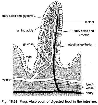
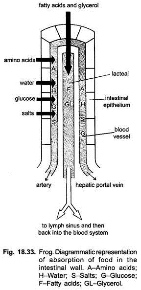
Absorption is that process by which the digested food is taken into blood. It takes place mainly in the duodenum and ileum as they are very much suited for this due to the development of various folds with villi-like processes, which increase the absorptive surface of these two regions of the alimentary canal. Each villus is richly supplied with blood capillaries and lymph vessel or lacteal. Water, mineral salts and other nutrients are directly absorbed through the mucosa.
The amino acids and glucose and fructose pass by diffusion from the mucosa into the blood capillaries of the intestine and finally reach the liver through the hepatic portal vein. The liver maintains the steady supply of the required amount of sugars and amino acids into blood. The excess sugar is stored as glycogen, whereas the excess amino acids cannot be stored in reserve but are converted into urea by liver cells which is eliminated as urine from the body by kidneys.
If sugar concentration falls from the normal in the blood, the reserve glycogen is changed into glucose by the liver cells and sent to the blood. From the blood the cells pick up the required amino acids to synthesise the proteins to form the protoplasm.
The glycerol and fatty acids pass into lymph vessels called lacteals. The glycerol can be absorbed easily as it is soluble in water, whereas the fatty acids as such cannot be absorbed as they are insoluble in water. So before absorption the fatty acids mix with bile salts to make themselves soluble so that they can be absorbed. After absorption in the lacteals, the glycerol and fatty acids are again converted into fat globules of much smaller molecules. Thus, fats enter as glycerol and fatty acids into the blood through lymph vessels.
Recent researches show that it is not necessary for all fats to be changed into glycerol and fatty acids for absorption, it is seen that fat droplets come in contact with villi and pass through their cell wall by pinocytosis into small lymphatic vessels.
(ii) Assimilation:
From the blood the different cells pick up required amount of various digested foods either to build new protoplasm or are used for providing energy. This phenomeon is known as assimilation. Vitamins convert the digested food into new protoplasm, mineral salts to form the parts of the protoplasm. Various foodstuffs under certain circumstances can be changed into other required substances.
5. Egestion of Indigested Food:
Digestion and absorption is completed in small intestine, whereas the indigested food enters rectum by peristalsis for storage and formation of faeces. Old epithelial cells, leucocytes, bile pigments and large number of bacteria form faeces which are removed from time to time through the cloacal opening.
