In this article we will discuss about the lymphatic system of frog with the help of suitable diagrams.
The blood is distributed to different parts of the body by the arteries. During its course of distribution certain portions of fluid plasma along with leucocytes escape into spaces (intercellular spaces) in the tissues through the capillaries as they are having very thin walls.
Most of the fluid is absorbed by blood capillaries but some of it passes by diffusion into special vessels called the lymph vessels. This squeezed fluid from the blood is known as lymph, while the special vessels are the lymphatic vessels, both constitute the lymphatic system.
The lymphatic system is an open system because it has lymph spaces between tissue cells and it does not form a complete circuit. Since lymph flows only in one direction, that is, from the tissues towards the heart, it has only capillaries and lymph vessels which are equivalent to veins.
ADVERTISEMENTS:
1. Lymph:
It is a colourless fluid which is a part of blood. Actually it is blood minus its red blood corpuscles and plasma proteins. It has less concentration of calcium and phosphorus than in blood. It has the power to coagulate slowly. It fills body spaces and bathes the tissue cells where it is known as tissue fluid. It is a medium of exchange between the blood and tissue cells. It carries food and oxygen to the cells and takes away water and other waste substances from the cells.
2. Lymph Vessels:
The lymph capillaries found between the tissue cells are interwoven with but independent of blood capillaries. Lymph capillaries form an anatomizing network in all the body organs except in the nervous system. The diameter of lymph capillaries is larger and less uniform than that of blood capillaries. At their commencement the closed ends of lymph capillaries have small swollen knobs which help in collecting lymph from tissues.
ADVERTISEMENTS:
The lymph capillaries unite to form larger lymph vessels or lymphatics which finally discharge their contents into veins. Thus, lymphatic’s are efferent vessels draining lymph into veins.
Lymphatics are made of connective tissue fibres with some unstriped muscles and are lined with endothelial cells. They are provided with valves present in pairs; they prevent backward flow of lymph.
3. Lymph Spaces:
ADVERTISEMENTS:
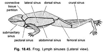
In frog in the tissues there are numerous lymph spaces from which arise lymph capillaries, the lymph capillaries unite to form lymph vessels, some of which dilate to form large lymph channels or sinuses which are as follows:
(i) Subcutaneous Lymph Sinuses:
They are large spaces found on the dorsal, ventral and lateral sides between the skin and muscles. They are separated from each other by fibrous membranes (septa).
(ii) Subvertebral Sinuses:
They are found around the dorsal aorta and also enclose the kidney.
(iii) Pericardial Sinuses:
They enclose the heart, their lymph is called pericardial fluid.
(iv) Coelom:
ADVERTISEMENTS:
It is a body cavity containing lymph which is commonly known as coelomic fluid.
4. Lymph Hearts:
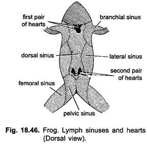
From the diffuse lymphatic system lymph is pumped back into veins by two pairs of lymph hearts. One pair is situated just behind the transverse processes of the third vertebrae or below the scapulae, opening into the subscapular veins. The second pair is found on either side at the end of the urostyle, they open into the femoral veins.
5. Spleen:
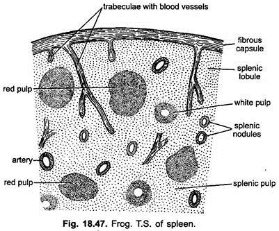
It lies near the rectum in the mesentery and is a dark red coloured body. It acts as a reservoir of blood, it destroys the worn out R.B.Cs., and produces antibodies, new R.B.Cs., and W.B.Cs., It is made of connective tissue and muscle fibres covered with fibrous capsule. It has many compartments containing splenic pulp (lymphatic tissue) which includes mainly the R.B.Cs., and W.B.Cs.
Blood:
The blood is really a fluid connective tissue containing fluid plasma in which red and white blood cells (corpuscles) are suspended. Plasma, of course, is non-living and constitutes about 60% of the blood, while blood corpuscles form the remaining 40%. Blood corpuscles are living cells.
1. Plasma:
Plasma is a liquid composed of 90% water and less than 6% of various inorganic and organic substances in solution. Inorganic substances are chloride, bicarbonates, sulphates, and phosphates of Na, K, Ca, Fe and Mg. Organic substances include plasma proteins like fibrinogen (coagulated substance), glucose, amino acids, hormones, enzymes and waste products. The amount of various constituents of plasma is fairly constant, though substances leave plasma all the time, and some others enter it.
2. Blood Cells or Corpuscles:
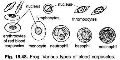
The blood cells or corpuscles include the red blood corpuscles (erythrocyte), white blood corpuscles (leucocytes) and thrombocytes, all are nucleated. None of these divides or multiplies in the blood. Certain of them are formed in the red bone marrow and others in the nodules of lymphatics.
(i) Red Blood Corpuscles or Erythrocytes:
In frog these are typically flattened cells of more or less elliptical shape but appear biconvex when seen along their edges. Each erythrocyte is yellowish in colour but when numerous are present they impart a red colour to the blood, therefore, they are popularly called red blood corpuscles. Each is filled with faint stain colour cytoplasm and contains a bulging biconvex nucleus.
They contain iron, water, various inorganic salts and respiratory pigment haemoglobin. Haemoglobin is a protein pigment which gives red colour to the corpuscles. It is a complex protein made of 90% globin (a colourless protein) and 5% hematin (red compound of iron).
The haemaglobin has a great affinity to oxygen, when it comes in contact, it becomes oxyhaemaglobin which breaks up into oxygen and haemaglobin in the tissue.
An erythrocyte is 23 x 14µ in size. Their number ranges from 250,000 to 450,000 per cubic millimeter of blood. In mammal they are circular, biconcave and non-nucleated and measure in their diameter. The life of erythrocytes is limited, in frog they live for about 100 days but in man for about 120 days.
(ii) White Blood Corpuscles or Leucocytes:
These are small nucleated and mostly amoeboid cells. Their number ranges from 5,000 to 7,000 per cubic millimeter. These are sometimes called phagocytes as they engulf bacteria and other foreign substances, and old tissue cells through the process of phagocytosis.
They have short life and can disintegrate in a day liberating their substances into blood. They are produced constantly in the liver, yellow bone marrow and spleen of frog but in mammals in red bone marrow, spleen, thymus and lymph nodes.
These are of two types depending upon the presence or absence of granules in their cytoplasm:
1. Granulocytes and
2. Non-Granulocytes.
1. Granulocytes or Polymorpholeucocytes:
These are of irregular shape having granular cytoplasm and lobed nucleus. These form 70% of total leucocytes and are produced in red bone marrow.
According to their staining property these are of three types:
(i) Basophils:
These cells are stained readily with basic dyes such as methylene blue or haematoxylin. They have a few large granules covering the two or three lobed nucleus. These are non-phagocytic and constitute 8% of the total leucocytes.
(ii) Eosinophils:
These are stained readily with basic dyes like eosin. They have many smaller granules and a nucleus of two lobes connected by a thread. These constitute about 3% of total leucocytes. These are also non-phagocytes.
(iii) Neutrophils (Heterophils):
These cells are stained with neutral dyes. They have numerous fine granules and a nucleus having many lobes. These form 67% of total leucocytes. These are phagocytic and engulf foreign bacteria and destroy them.
2. Non-Granulocytes:
These have a non-granular cytoplasm and are produced from the special cells in the lymphatic nodules. These are extremely variable in size.
These are of two types:
(i) Lymphocytes and
(ii) Monocytes.
(i) Lymphocytes:
These are small in size, rounded and have a large nucleus with an indentation. They have small amount of cytoplasm which surrounds the nucleus. These form 28% of total leucocytes.
(ii) Monocytes:
These are larger having large excentric nucleus indented on one side. They have more cytoplasm than in lymphocytes. They are phagocytes and are produced in spleen or lymph nodules.
All leucocytes exhibit their chief activities in tissue cells, they transport substances, remove dead cells and engulf bacteria. Whenever bacteria or foreign particles enter the body or any part is ingested, the phagocytic neutrophils and monocytes squeeze out from the endothelium to engulf the bacteria and foreign particles. These cells produce antibodies to neutralise the infection caused by bacteria. The foreign particles or substances which cause the production of antibodies are called antigens.
3. Thrombocytes (Spindle Cells):
These are small spindle-shaped nucleated cells which are known as thrombocytes.
In mammals instead of thrombocytes blood platelets are found which are round, colourless, biconvex and non-nucleated.
Clotting of Blood:
Clotting of blood is its most important property due to which blood is never escaped from the body as it clots and seals the site of injury. Blood moving normally through the circulating system does not clot because of the presence of antithrombin, an organic compound called heparin produced in liver.
During clotting the fibrinogen protein present in the plasma of blood is precipitated by the action of enzyme thrombin to form a dense meshwork of fine threads of fibrin in which various types of corpuscles become entangled. This results in the formation of clot. Thrombin does not occur in an active form in the blood but it occurs as a precursor called prothrombin.
Prothrombin in the presence of calcium ions is converted into active thrombin. When a blood vessel is injured a substance called thromboplastin or thrombokinase is released from the disintegrating thrombocytes (blood platelets) and from the injured tissues.
This substance neutralises the effect of hepagin, and thrombin causes the precipitation of the fibrinogen proteins into fibrin. It is, thus, apparent that clotting of blood depends on the presence of prothrombin, thromboplastin and calcium ions.
Functions of Blood:
The general functions of the blood can be summarised as follows:
1. Transport of Oxygen:
Blood contains haemoglobin in its erythrocytes which with oxygen forms oxyhaemoglobin and transports it to different parts of the body for the oxidation to release energy.
2. Removal of Carbon Dioxide:
After the oxidation of food material in the tissue cells, carbon dioxide is formed which is picked up by plasma and erythrocytes which take it to the lungs or skin from where it is released.
3. Transport of Food Materials:
All the digested food materials are absorbed through the intestinal wall and find their way into the blood and are carried in the plasma until they are taken by the tissue cells.
4. Removal of Unwanted Substances (Metabolic Wastes):
Plasma of the blood picks up all the metabolic wastes like urea and uric acid from various parts of the body and takes them first to the liver and then to the kidneys from where these are eliminated.
5. Transport of Other Substances:
There are many other substances such as hormones and antitoxins which are also transported by the blood to different parts of the body in order to bring harmonious cooperation and integration of various parts of the body.
6. Clotting of Blood:
It prevents loss of much blood from an injury by forming a clot from its fibrinogen over the site of injury.
7. Equilibration of Water Content:
It maintains the equilibration of water contents in the tissue cells.
8. Regulation of Body Temperature:
It regulates and equalises body temperature by carrying heat away from the region where it is produced.
9. Protection from Diseases:
Phagocytic leucocytes combat bacteria and foreign bodies, thus, prevent diseases. The plasma contain antitoxins and antibodies which neutralise the harmful effects of bacteria and toxins.
10. Healing of Wounds:
Blood brings all the essential substances to the site of injury for healing wounds.


