In this article we will discuss about:- 1. Meaning of Pituitary Gland 2. Development of Pituitary Gland 3. Location 4. Anatomy.
Meaning of Pituitary Gland:
The hypophysis or pituitary gland is a small gland near the brain and is an essential link in the neuroendocrine system. It has little capacity to function independently though the gland has often been referred to as the master endocrine gland. This importance may have given because it controls several other endocrine glands and no part of the body is exempt from its influences.
Since, it is subservient to the nervous system and some other endocrine glands; it is misleading to refer to it as the “master gland” of the body.
Development of Pituitary Gland:
The entire pituitary gland is ectodermal in origin. Its posterior lobe or neurohypophysis originates from the infundibulum of the brain, an out pouching of the hypothalamus. The anterior lobe or adenohypophysis originates from Rathkes’ pouch, an ectodermal evagination of the oropharynx and migrates to join the neurohypophysis.
ADVERTISEMENTS:
The portion of Rathkes’ pouch in contact with the neurohypophysis develops less extensively and forms the intermediate lobe. This lobe remains intact in some species but in human, its cells become interspersed with those of the anterior lobe.
Location of Pituitary Gland:
Beneath the hypothalamus, near the optic chiasma, the pituitary gland lies at the base of the skull in a portion of the sphenoid bone called the sella turcica.
Anatomy of Pituitary Gland:
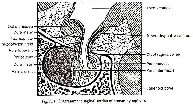
The pituitary gland is a compound gland with internal secretion. It is encapsulated by the Dura mater; a shield-like fold of this membrane forms the diaphragm sellae and extends around the infundibular stalk.
The pars tuberalis, a part of adenohypophysis is a thin epithelial plate of cells that in adult may surround the infundibular stalk and extend some distance below the tuber cinereum. In young human, a narrow band of tissue, the pars intermedia is found between the pars distalis and the neural lobe. In adults, this lobe merges with the neural lobe and tends to become obscure.
ADVERTISEMENTS:
The pituitary gland receives its blood supply from the superior and inferior hypophysial arteries. The pars distalis is vascularized by hypophyseal portal vessels that arise from the capillary beds within the median eminence of the hypothalamus. The pars nervosa receives a separate blood supply from the inferior hypophyseal artery.
The pars intermedia, if present is relatively avascular. It is generally assumed that all hormones produced by the adenohypophysis are released directly into efferent portal veins to be carried through the systemic circulation to distant target tissues.
The nerve supply of the hypophysis consists of sympathetic fibres from the surrounding perivascular plexus, parasympathetic fibres from the petrosal nerves, and the hypothalamo-hypophyseal tract. However, only the pars tuberalis receives the sympathetic nerves but none in the pars distalis. Since pars tuberalis secretes no hormones, the nerve fibres that end there, are vasomotor rather than secretomotor.
The pars nervosa has a conspicuous feature – a large number of secretory axons terminating there. The perikarya of these axons are located in the para-ventricular and supra optic nuclei in mammals (Fig. 7.11). The fibres sweep down the pituitary stalk and are grouped into supraoptico- and tubero-hypophyseal tracts. Some of these fibres are known to terminate in the median eminence and in the pars intermedia.
Different Parts of Pituitary Gland:
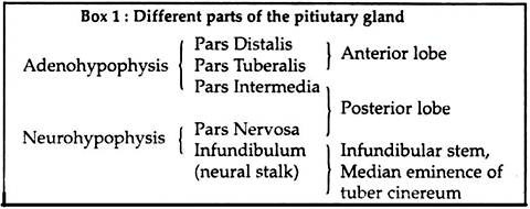
The human pituitary is composed of an adenohypophysis (glandular or epithelial hypophysis) and a neurohypophysis. A variety of anatomical designations have been applied to the pituitary but the most widely accepted terminology is provided in Box 1.
A. Adenohypophysis:
1. Pars distalis:
The pars distalis of adenohypophysis is composed of irregular masses and cords of epithelial cells separated by sinusoids and supported by a loose framework of connective tissue. Routine staining of this region with stains like eosin and hematoxylin or with Mallory aniline blue or azan reveals two major types of gland cells.
The chromophils containing characteristic cytoplasmic granules and are generally considered to be secretory. The other type is chromophobes that do not possess conspicuous secretory granules. Most cytologists considered these chromophobes as the progenitors of other cell types or as de-granulated, resting stages of chromophils.
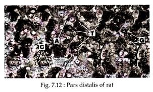
On the basis of tinctorial responses, chromophils are further classified into acidophils (Fig. 7.12A) and basophils, though cytoplasmic granules of these cells often respond to both acid and basic stains. Later on, a more specific scheme for designating hormone-secreting cells of pars distalis has evolved on the basis of data obtained mainly by electron microscopy and immunocytochemical techniques.
The cell types of pars distalis according to the scheme are:
ADVERTISEMENTS:
(a) Somatotrophs:
The growth hormone secreting cells are called somatotrophs. These are acidophilic in routine staining (Eosin-hematoxylin) and are located in the lateral portion of the adenohypophysis. These middle-sized ovoid cells account for about 50% of pars distalis and are PAS- (periodic acid-Schiff stains) negative.
The majority of these cells have spherical nucleus and well developed lamellar rough endoplasmic reticulum (RER), located at the periphery of the cells. A prominent Golgi complex with a few secretory granules is very common to these cells. Granule size (by electron microscopy) varies from 150 to 600 nm in diameter.
Apart from variations in secretory granule size and morphology, a contingent of GH-cell population is capable of producing prolactin as well. These dual secretors, called mammosomatotrophs are considered transitional cells with potential for alternating between somatotroph and lactotroph functions.
(b) Lactotrophs:
These prolactin secreting cells are also known as mammotrophs or luteotrophs. These cells belong to the acidophil cell series and are less numerous than the somatotrophs, constituting about 10-30% of total cell population. These cells remain scattered throughout the pars distalis.
Under electron microscope two types of lactotrophs are shown. The densely granulated ovoid mammotrophs contain parallel arrays of well-developed RER and a prominent Golgi complex. The spherical and ovid granules of these cells are numerous and measure 400-700 nm. These cells are uncommon.
The common prolactin cells are middle sized or small polyhedral cells with long processes. The lucent cytoplasm contains abundant RER, a well-developed Golgi complex and small (200-350 nm) sparse secretory granules. The number of prolactin cells increases sharply during pregnancy and lactation and as a result of estrogen treatment.
(c) Thyrotrophs:
Thyrotrophs are the thyroid-stimulating hormone producing cells. These cells are basophilic in nature because of their glycoprotein content. These cells constitute about 5% of pars distalis (Fig. 7.12T). These cells are located in the anteromedial and anterolateral portions of the gland.
Electron microscopy reveals that the elongated angular cells possess short profiles of slightly dilated RER and globular Golgi complex with numerous vesicles. The small secretory granules measure 100-300 nm, are mostly Spherical, vary in electron opacity and are often positioned near plasma-lemma. During various states of hypothyroidism, these cells undergo hypertrophy and hyperplasia.
(d) Corticotrophs:
Corticotrophs are adrenocorticotrophic hormone (ACTH) producing cells that constitute about 20% of adenohypophysial cells. These cells are basophilic but are often referred to as chromophobes (Fig. 7.12C) and PAS-positive. By electron microscopy, the elongated, angular cells appear to have round or slightly irregular nuclei.
The cytoplasm has well-developed, widely dispersed RER and prominent Golgi with immature secretory granules. Cytoplasm also harbors a large number of spherical or irregular or heart-shaped secretory granules having different electron density and measuring 250-400 nm in diameter.
In states of glucocorticoid excess, corticotrophs undergo degranulation and a micro-tubular hyalinisation known as Crookes’ hyaline degeneration.
(e) Gonadotrophs:
According to current views, a single type of cell secreting both gonadotrophic hormones (follicle-stimulating hormone or FSH and luteinizing hormone or LH) is known as gonadotrophs. These cells constitute about 15% of pars distalis cell population and are located throughout the entire anterior lobe.
They are PAS-positive and basophilic (Fig. 7.12G). Under electron microscope, these ovoid cells appear to have a considerable surface area adjoining the capillaries. The cells have spherical or ovoid nuclei. Cytoplasm contain well-developed RER, rod-shaped mitochondria with numerous lamellar cristae and a large ring-shaped Golgi complex.
The majority of secretory granules measure 250-300 nm but large granules with a diameter of 400-450 nm may also occur. The gonadotrophs become hypertrophied during states of primary gonadal failure such as menopause, Klinefelter’ syndrome and Turner’s syndrome.
2. Pars tuberalis:
This thin sleeve-like region of adenohypophysis is only 25-60 µm in thickness (Fig. 7.11), with its thickest portion on its anterior aspect. It is separated from the infundibular stalk by a thin layer of connective tissue that is continuous with the pia arachnoid and a similar layer covers the outer surface.
The distinctive morphological feature of pars tuberalis is the longitudinal arrangement of its cords of epithelial cells, which occupy the interstices between those longitudinally oriented blood vessels. Its principal cells are 12-18 µm in size and are cuboidal or low columnar in form.
Cytoplasm of these cells contains short rod-like mitochondria, small dense granules, numerous lipid droplets, occasional colloid droplets and significant amount of glycogen. The cells may form follicle like structures.
Islands of squamous epithelial cells may also be found. Although some of the cells appear to contain secretory granules, no specific hormone has been identified and thus the function of the pars tuberalis remains unknown.
3. Pars intermedia:
This lobe is rudimentary in adult human, but in other vertebrates like rat and in poikilotherms, the intermediate lobe is composed of polygonal cells that take basic dyes and in which secretory granules with diameter of 200-250 nm have been observed. Some of the granules are electron-dense; others are pale, owing to extraction of some of their substances during specimen preparation.
In addition, cytoplasm of these cells also contains well, developed ER and Golgi complex. In some species, vesicles containing colloid are present in these cells. Cells of pars intermedia secrete melanocyte- stimulating hormone (MSH). In frog and elasmabranchs, two types of nerve endings have been found in the pars intermedia of adenohypophysis.
B. Neurohypophysis:
Neurohypophysis consists of median eminence of tuber cinereum, the infundibular stem and pars nervosa. The pars nervosa portion of neurohypophysis consists of branching cells, called pituicytes (P), thick network of fine un-myelinated nerve fibres that are the termination of the hypothalamohypophyseal tract (Fig 7.13).
In histological sections of the posterior lobe stained with alum hematoxylin, aggregations of deeply stained material of varying size are found in dilations of the axons throughout the neural lobe. These were formerly called Herring bodies (H). In electron micrographs, they are now found to be large aggregations of small neurosecretory granules.
It is estimated that each axon of the hypothalamohypophyseal tract has about 450 expansions along its course containing, on an average, over 2,000 neurosecretory granules. About 60% of the neurosecretory material resides in these axonal dilations and 40% in the axon endings that are associated with fenestrated capillaries throughout the neural lobe.
The axon terminals of neurohypophysis are distinguishable from other axonal dilations by the presence of numerous small vesicles, in addition to the secretory granules.
The organisation of pars nervosa cannot be adequately described without inclusion of the cell bodies that are located beyond its boundaries. The cell bodies are present in the supraoptic nucleus located above and lateral to the optic chiasma and in the para-ventricular nucleus at a higher level in the lateral wall of third ventricle.
Un-myelinated axons of neurons of these cells form the hypothalamohypophyseal tract that descend into, and make up the bulk of the pars nervosa.
The pituicytes with slender processes are joined to the processes of neighbouring pituicytes or stellate cells to form a three dimensional network among the axons of hypothalamic neurosecretory cells. In human, the pituicytes are highly variable in size and shape and contain lipid droplets and depositions of lipochrome pigment.
The processes of pituicytes often intimately en-sheath the terminal dilations of the axons. Such topographical relationship of pituicyte to the axons is similar to that of neuroglial cells of the central nervous system, though no physiological interaction has yet been established for such relationship.
Most workers suggest that the hormones of neurohypophysis are not secreted by pituicytes, but are produced by certain neurosecretory cells of hypothalamic nuclei.
The secretions move along the axons of hypothalamohypophyseal tract into the neural lobe, where it is discharged, stored, possibly modified and released into the general circulation as needed. Thus, the neural lobe is a depot for the storage of hormones.


