In this article we will discuss about Flagellar Locomotion in Euglena:- 1. Introduction to Flagella in Euglena 2. Structure of Flagellum in Euglena 3. Ultrastructure 4. Role.
Introduction to Flagella in Euglena:
A common plan of organization in the non-muscular contractile system of animals is found both in flagella and cilia. These structures with certain associated fibrillar systems, provide organelles of movement not only for different protozoa, but also in many metazoan animals where that function as an important effector structure.
The effect of flagella upon the movement of a protozoa is best exemplified by Euglena — an organism, 55-100 µm in length, found swimming freely on the surface of fresh water bodies like pond, canal, lake etc.
Structure of Flagellum in Euglena:
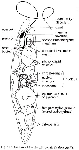
In general, flagellum is a long whip like organ which protrudes to the exterior from the cell body and permits mechanical work without any marked change in the form of the effector cell. In Euglena, there are two flagella. One of them is equal in length to body while other is short. They emerge out through the gullet — a narrow depression at the exterior end of the spindle-shaped body.
ADVERTISEMENTS:
The gullet leads to a flask-shaped non-contractile reservoir (Fig. 2.1). The flagellum bifurcates into two at the middle of the reservoir. These two flagella originate from two compact basal granules or blepharoplasts, situated in the cytoplasm just beneath the base of the reservoir.
In most species of Euglena, the two flagella originate separately from two blepharoplasts and the shorter one soon after its emergence unites with the longer one (Fig. 2.1).
Ultrastructure of Flagellar in Euglena:
Electron microscopy has shown that the long flagellum in Euglena has two parts:
ADVERTISEMENTS:
1. Outer Coat:
It is a contractile membranous sheath that is continuous with the cell membrane.
2. Axoneme:
It is the inner core, composed of microtubules and other proteins. Microtubules are normally long, hollow tubes formed of two types of proteins viz., a tubulin and p tubulin.
ADVERTISEMENTS:
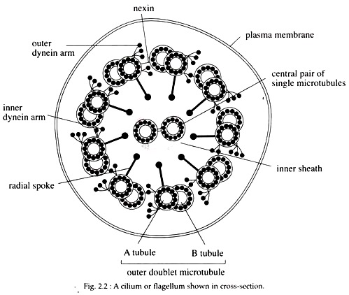
In the axoneme, the microtubules are modified and arranged in a ring of nine special doublets of microtubules surrounding a central pair of single microtubule (Fig. 2.2). This “9 + 2” array is the characteristic of axoneme of almost all forms of cilia and flagella.
This microtubules extend continuously throughout the length of axoneme. In the centre, the pair of single microtubules are complete microtubules, while in the outer ring, each doublet is composed of one complete and one partial microtubules known as the A and B tubules respectively. Each doublet in the outer ring is provided with sets of arms that join neighbouring doublets.
Each arm is composed of a protein called dynein. Pairs of inner and outer arms are spaced all along each A tubule at regular 24 nm intervals. The outer doublets are connected circumferentially by another protein called nexin links at intervals of about 96 nm. A series of radial spoke with a periodicity of 88 to 96 nm extends from the A sub-tubule to the central pair of microtubules (Fig. 2.2).
All flagella arise from a basal body. When the basal bodies are distributed to daughter cells during mitosis, they typically arrange themselves at each pole of the mitotic spindle and are then designated as centrioles. A region around the basal bodies and centrioles, called the microtubule organizing centre, controls the above ‘mentioned organized assembly of microtubules.
The ultrastructure of the basal bodies is like that of an axoneme except that the central singlet are absent and the nine fibrils in the outer circle are triplets, two of these being continuous with the doublets of the flagellum. Dynein arms however, are absent in the triplets.
Role of Flagella in Locomotion:
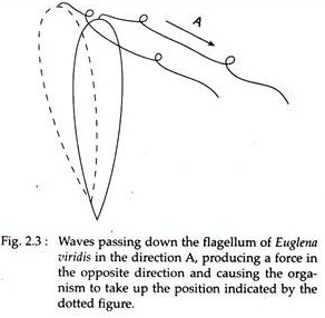
In Euglena, the movement of flagella commonly involves the generation of waves that are transmitted along it, either in a single plane or in a corkscrew pattern. The waves arise at the base of the flagellum, from the wall of the reservoir, apparently by two roots. The waves then pass to tip of the main flagellum, which beats at a rate of about 12 strokes per second and also shows a movement of rotation.
This rotation causes the tip of the organism to rotate (Fig. 2.3), while at the same time pushing it to one side (Fig. 2.4). Because of this, Euglena rotates as it swims (at a rate of about 1 turn per second) and it also follows a corkscrew course (Fig. 2.4).
ADVERTISEMENTS:
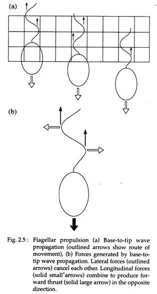
The movement of its body is thus comparable with that of propeller, for it sets up forces on the water that bring about forward displacement. When an undulation moves along the flagellum, it also generates lateral forces. These forces are usually symmetrical, the left-directed forces cancel the right directed forces, and only the longitudinal force remains to move the cell forward (Fig. 2.5a & 2.5b).
The relationship of flagellar ultrastructure to movement has received much attention in recent years and the sliding tubule model is now widely accepted. According to this theory, the movement of a flagellum is produced by the bending of the core or axoneme. The bending force is produced due to active sliding of adjacent outer doublets against each other.
In the presence of ATP, the dynein arm on one doublet attaches to the adjacent doublet and flexes, causing the doublets to slide past each other by one increment. Successive attachments and flexes cause the doublets to slide smoothly past one another over a distance sufficient to bend the flagellum.
If a flagellum is severed from a cell by a laser beam, the isolated structure continues to propagate bending movements in a normal way, indicating that the motile machinery is contained in the axoneme itself and its movement do not depend on a motor at its base.
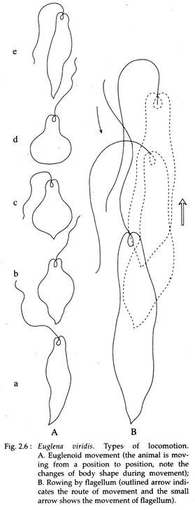
Sometimes, Euglena shows a very peculiar motion in which waves of contraction pass along the body from anterior to posterior end and the animal creeps forward. This contraction is brought about by the stretching of protoplasm on the pellicle or by localised fibrils called myonemes in the cytoplasm.
This type of locomotion is known as Euglenoid movement (Fig. 2.6A). An Euglena can also move by rowing. During rowing, the flagellum is held rigid and is slightly arched in the direction of the stroke. In recovering the position, it bends as it is drawn back so as to face minimum resistance (Fig. 2.6B).
