In this article we will discuss about the Nervous System of Various Invertebrates:- 1. Nervous System in Cnidarians 2. Nervous System in Asterias 3. Earthworm 4. Cockroach 5. Pila.
Contents:
- Nervous System in Cnidarians
- Nervous System in Asterias
- Nervous System of Earthworm
- Nervous System of Cockroach
- Nervous System of Pila
1. Nervous System in Cnidarians:
The cnidarian nervous system is basically a plexus of relatively short-fibered bipolar and multipolar nerve cells, together with neurites from sensory cells. In Hydra, there are two nets — a main plexus between the epidermis and the musculature and another is less highly developed network associated with the gastro-dermis and connected at various points with the epidermal plexus.
ADVERTISEMENTS:
These nerve nets are recognized as a sympathetic association of neurons in which fibres make contact but do not fuse. The neurons are more concentrated in the hypostome and the pedal disc. Here the plexus has a circular arrangement, suggesting the appearance of nerve ring.
2. Nervous System in Asterias:
The nervous system of Asterias is very simple. It includes only nerve net and has no central ganglionic formation. The nervous system consists of three systems: oral or ectoneural system, deep or hypo-neural system and coelomic nervous system (Fig. 1.135).
Oral or Ectoneural Nervous System:
ADVERTISEMENTS:
This system forms the main part of the nervous system and is situated below the epidermis. It includes a nerve pentagon situated in the peristomial membrane surrounding the mouth. The nerve pentagon at each radius gives off a radial nerve which runs as a slender thick band throughout the ambulacral groove.
The radial nerve is situated just below the radial canal as median integumentary thickening. The radial nerve ends as a sensory pad or cushion at the base of the terminal tentacles. The radial nerve gives branches to the tube-feet and becomes continuous with the sub-epidermal nerve plexus of the body wall.
The nerve pentagon and the radial nerves consist of nerve fibrils with scattered bipolar and multipolar nerve cells. The radial nerve appears like a thick ‘V’- shaped body below the hypo-neural sinus. It is separated from the hypo-neural sinus by a thin dermis and coelomic epithelium.
Deep or Hypo-neural Nervous System:
ADVERTISEMENTS:
The different nerves composing this part are motor nerves. This nervous system comprises of Lange’s nerves. Lange’s nerve is a plate of nervous tissue situated beneath the coelomic epithelium of the hypo-neural sinus and forms a lateral lining on the wall of this sinus.
Lange’s nerve extends as five inter- radial nerve thickenings above the main nerve pentagon of the ectoneural system. Each Lange’s nerve innervates the muscles of the corresponding arm.
Coelomic Nervous System:
The sub-epithelial nerve plexus at the outer ends of the ambulacral ossicles forms marginal nerve cord on two sides of the arms. The marginal nerve cord gives off lateral motor nerves which proceed to the aboral side between the adambulacral and ambulacral ossicles to reach the coelomic lining. Beneath the entire coelomic lining the nerves form plexus which innervates the body wall musculature and the gonads.
One important feature that develop in the nervous system of star fish is the small areas of neuropile. In the neuropile, the nerve fibres show close inter weaving instead of the regular parallel alignment. The fibres within the neuropile contain numerous vesicles of diameter 35 mm to 100 mm.
These vesicles are supposed to contain chemical transmitter substances. The neuropile areas provide the transmission of signals from the sensory fibres to those of the interneurons and motor cells. This is an important advancement towards the development of separate conducting systems and the beginning of a central nervous system.
3. Nervous System of Earthworm:
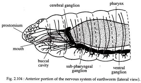
The nervous system comprises of a central nervous system (Fig. 2.104) from which peripheral nerves are given to the different parts of the body and the receptor organs.
Central Nervous System:
ADVERTISEMENTS:
It consists of a pair of closely packed supra-pharyngeal ganglia or cerebral ganglia forming the brain and situated in the third segment of the body above the pharynx. From the brain, nerves are given off to the prostomium and walls of the buccal chamber. Two loops called circum pharyngeal connectives arise, one from each ganglion of the brain.
They encircle the pharynx and meet with a pair of sub-pharyngeal ganglia lying below the pharynx and in the anterior part of the fourth segment. The connectives give off nerves to the first segment of the body and to the buccal chamber and the sub-pharyngeal ganglia give branches to supply the second, third and fourth segments.
A ventral nerve cord extends from the sub-pharyngeal ganglia to the last segment of the body and runs along the mid-ventral line.
The nerve cord, though appears single, is in reality made up of two cords. Behind the fourth segment the cord presents a swelling in each segment. Each swelling is formed by fusion of paired ganglia. From each of these ganglia, three pairs of peripheral nerves are given to the body wall and viscera. These nerves contain both afferent and efferent fibres.
Receptor Organs:
The structures which receive stimuli and convey the same to the central nervous system are called receptors.
The receptors found in the body of earthworm are described below:
(a) Epidermal Receptors:
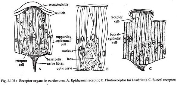
These receptors are placed in the epidermis as groups of long slender receptor cells (Fig. 2.105A) which are tactile in function.
(b) Photoreceptors:
Each photoreceptor (Fig. 2.105B) is a single cell with a distinct nucleus and an optic organellae. These receptor cells are housed in the epidermis. The cells are numerous at the anterior end of the body. The number gradually decreases posteriorly. The last segment may even contain one such cell. The receptor cells are absent on the ventral surface.
(c) Buccal Receptors:
These receptors are group of cells placed in the buccal cavity in large numbers. These receptors are concerned with chemical stimuli and serve in smelling and tasting food particles (Fig. 2.105C).
4. Nervous System of Cockroach:
The nervous system of cockroach includes:
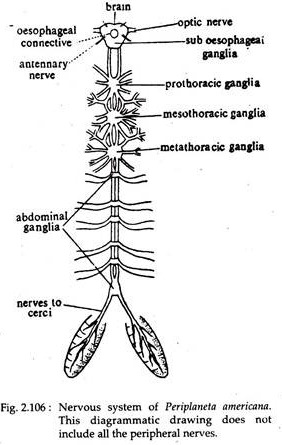
(a) Central Nervous System (Fig. 2.106),
(b) Peripheral Nervous System,
(c) Sympathetic Nervous System and
(a) Central Nervous System:
The central nervous system consists of several specialised parts which control the working of the different structures of the body through the peripheral nervous system.
Each of the specialised parts of the central nervous system are discussed below:
i. Brain or Supra-oesophageal Gunglia:
These are paired and large ganglia, present in the posterior side of the head region and on the dorsal side of the oesophagus. Each ganglion consists of a hilobed protocerebrum, a deutocerebrum and tritocerebrum.
ii. Sub-oesophageal Ganglion:
This ganglion is present in the mid-ventral region of the head and just ventral to the oesophagus. It is formed by the fusion of two ganglia.
iii. Circum-oesophageal Connectives:
These are short and broad, paired nerves which originate one from each supra-oesophagus to unite with the sub-oesophageal ganglion. The two connectives are connected by one large and one small transverse commissure.
iv. Ventral Nerve Cord:
It is formed by two solid nerves, which begin from the posterior end of the sub-oesophageal ganglion and run posteriorly along the mid-ventral line of the body cavity. In each thoracic segment it bears a prominent ganglion and in the abdomen there are six abdominal ganglia. First four abdominal ganglia are present, one in each of the first four abdominal segments.
The fifth abdominal ganglion is present near the junction of’ fifth and sixth segments. The sixth abdominal ganglion denotes the termination of ventral nerve cord. It is twice the size of other abdominal ganglia. This last abdominal ganglion is present more or less on the eighth segment.
(b) Peripheral Nervous System:
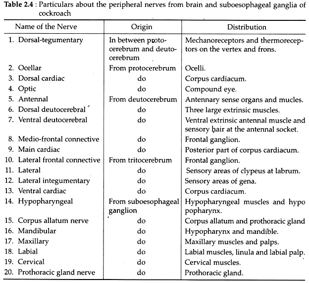
These nerves are given out from the ganglia of the central nervous system. The name and distribution of different peripheral nerves which are given from the brain and sub-oesophageal ganglia are shown in the table (Table 2.4).
Each thoracic ganglion has eight pairs of nerve roots. With the first pair the ventral nerve cord communicates. The remaining seven pairs of thoracic nerves supply the muscles of the thoracic segment, legs and wings. The metathoracic ganglion in addition sends peripheral nerves to the first abdominal segment.
Each of the first five abdominal ganglia sends out pair of stout nerves to supply different parts of the abdominal segment posterior to it. The sixth abdominal ganglion sends out seven to eight pairs of nerves to the different structures present in the seventh, eighth, ninth and tenth abdominal segments.
(c) Sympathetic Nervous System:
It is represented by a slender ganglionated sympathetic nerve, which begins from circum- oesophageal connectives to innervate the involuntary muscles of the alimentary canal.
(d) Sense Organs:
The sense organs of cockroach may be grouped according to their functions as mechanoreceptors, photoreceptors, thermo-receptors and chemoreceptors. In addition, other kind of receptors, for responding to humidity changes, are recorded in the cockroach, Blatella. These are called hygro-receptors.
Mechanoreceptors:
Following sensory structures are considered as mechanoreceptors because they are concerned with the reception of stimuli in the form of touch, pressure, vibration and air current.
(a) Cuticular Hair:
These are present in the cuticle either in the form of bristles, spines, plates or collection of hair. Tactile bristles are present in the cercus and are extremely sensitive to touch. Large spines on the femur and tibia of leg perform similar function.
A collection of hair, known as hair plales, are present on certain parts of the body for receiving stimuli in the form of touch. A group of specialised hair which are lodged in sockets occur in the cercus for receiving air movements ‘and low frequency sounds. These are called auditory hair.
(b) Companiform Stress Receptors:
They are present as thick ridges on the segments of the leg, on the labial palps and its adjoining areas. These receptors are believed to respond to different pressure on the cuticular surface.
(c) Chordotomal Organs:
These specialised sense organs meant for receiving vibrations are distributed on the legs. These are aggregates of specialised cells which remain arranged either parallelly or in the shape of a fan.
Photoreceptors:
The eyes and ocelli are the two important photoreceptors of cock roach, in addition to the general surface of the body. These can also detect light stimuli through cuticular receptors.
(a) Compound Eyes:
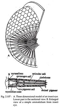
Two compound eyes are situated on dorsolateral sides of the head of cockroach. The external surface of the eye is covered by a transparent cuticle. Each eye is composed nearly of 20,000 visual units, each one of which is called ommatidium (Fig. 2.107).
Each ommandium consists of following structures:
(i) Cornea:
It is the outermost transparent cuticular layer which gives the outermost part of the eye a honeycomb-like appearance (Fig. 2.107B).
(ii) Corneagen Cells:
Immediately beneath the cornea a pair of corneagen cells is present which are responsible for the replacement of cornea (Fig. 2.107B).
(iii) Crystalline Cone:
It is a well-developed triangular transparent body surrounded by cone cells. It works as lens (Fig. 2.107B).
(iv) Cone Cells:
These cells encircle the cone and provide nourishment to the lens,
(v) Rhabdome:
Elongated and transversely striated body, which extends up to the border of the cone (Fig. 2.107B).
(vi) Retinular Cells:
These elongated sickle-shaped cells secrete the rhabdome and encircle it to provide nourishment,
(vii) Pigment Sheath:
Iris and retinal pigment sheaths together separate one ommatidium from another. Thus only mosaic type of image formation occurs.
(b) Ocellus:
This is also known as simple eye. It is present near the base of antenna as a white area. It consists of outer flattened corneal cells, 3-5 retinular cells and one rhabdome formed by the inner margins of the retinular cells.
It was formerly believed that these ocelli are of uncertain functions. Recently the light detecting ability of these structures has been proved. It has been demonstrated that cockroaches can respond to the change of light even when the compound eyes are painted. But it fails to do so if the ocelli are covered.
Temperature Receptors:
The temperature receptive sense organs are present in pads between the first four tarsal segments of the leg.
Chemoreceptors:
These receptors are responsible for detecting chemical stimuli in the form of smell and taste. The long and annulated antennae are beset with two types of sensory structures — thick-walled bristles and thin-walled hairs.
These bristles and hairs are responsible for the perception of smell. The tips of the maxillary and labial palps, inner surfaces of the mouth parts and the inner border of the mouth and pharynx possess sensory structures for contact, chemoreception and gustatory responses.
5. Nervous System of Pila:
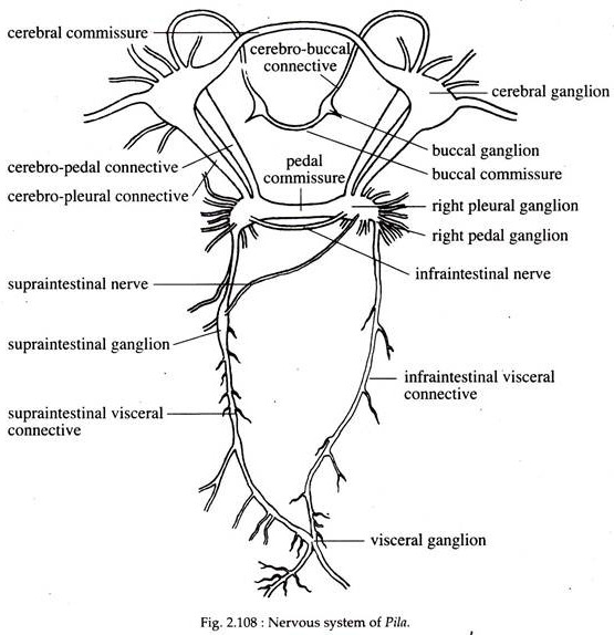
The nervous system consists of ganglia; commissures, connectives and the nerves to different organs (Fig. 2.108).
Ganglia:
The main ganglia are:
(a) One pair of roughly triangular cerebral ganglia situated on the dorsolateral sides of the buccal mass, one on each side of the head.
(b) One pair of pleuropedal ganglia placed below the buccal mass on the lateral sides. Each pleuropedal ganglionic mass is formed by the fusion of pleural and pedal ganglia. The infra-intestinal ganglion is also fused with the right pleuropedal mass.
(c) Visceral ganglion is very large and appears to be unpaired. It is a bilobed structure and is formed by the fusion of two separate ganglia. The visceral ganglion is placed posteriorly very close to the heart.
(d) A pair of buccal ganglia are situated on the buccal mass on the two sides of the oesophagus.
(e) A single supraintestinal ganglion is located near the middle of the left pleuro- visceral connectives.
Commissures:
The nerve connections between two similar ganglia are called commissures. The ganglia are placed on the opposite sides of the body. Two cerebral ganglia are connected by a thick nerve cord called cerebral commissure. The buccal ganglia are also connected by a delicate buccal commissure. The inner sides of the pleuropedal ganglia are connected by a broad nerve called pedal commissure.
Connectives:
The nerve connections between two dissimilar ganglia are called connectives. The ganglia may be situated on the same or opposite sides of the body. The cerebral ganglia and the buccal ganglia are connected by cerebrobuccal connectives. The pleuropedal ganglia are connected on each side with the cerebral ganglion by cerebropedal and cerebropleural connectives.
That the pleuropedal ganglia are formed by the fusion of separate pleural and pedal ganglia is indicated by the presence of an indistinct constriction and the existence of two separate connectives joining the cerebral ganglia. The pleural ganglion is connected with the visceral ganglion by a pleurovisceral connective on each side.
The right pleurovisceral connective lies below the level of the oesophagus and is generally designated as infraintestinal visceral connective and the left pleurovisceral connective is situated above the level of the oesophagus and is termed as supraintestinal visceral connective.
The supraintestinal ganglion is connected with the right pleuropedal ganglion by an oblique nerve placed above the oesophagus called supraintestinal nerve. A very slender nerve called infraintestinal nerve is present connecting the two pleural ganglia of two sides.
Nerves to the different parts of the body:
Each cerebral ganglion innervates the eye, the snout and the tentacles on its side. The statocyst is also innervated by a slender nerve arising from the cerebral ganglion. The pedal ganglion gives out numerous nerves to the foot and the pleural ganglion supplies the mantle.
The supraintestinal ganglion supplies nerves to the ctenidium and the pulmonary sac. The visceral ganglion sends nerves to kidney, genital organ, pericardium and intestine.
The buccal ganglion innervates the buccal mass:
Chiastoneury:
The nervous system of Pila exhibits streptoneurons chiastoneury condition. This is the result of torsion of visceral mass which has made the whole nervous system asymmetrical. The complexities in the nervous system in Pila are due to complete migration of the anal and genital openings in the oral end.
The chiastoneury is not so clear in Pila and typical figure of 8-like arrangement is not produced between the supra- and infra-intestinal nerves. No crossing is possible on the right side because of the shifting and fusion of infra-intestinal ganglion with the right pleural ganglion.
The zygoneury (a secondary connection between pleural and supraintestinal ganglia) is present only on the left side. So the typical chiastoneurous condition with double zygoneury as seen in many gastropods is not clear in Pila.
Sense Organs:
The sense organs are quite well developed.
Osphradium:
The osphradium is the organ for taste. It helps to taste the chemical and physical qualities of the incurrent water and also assists in the selection of food. There is only one osphradium which remains suspended from the roof of the pallial cavity on the left side.
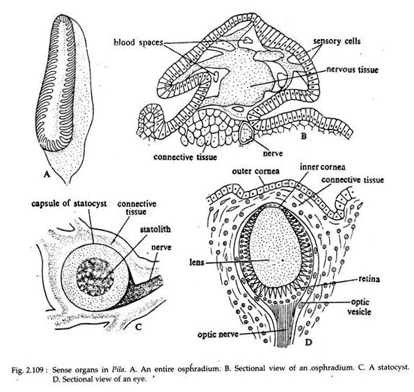
It is small in size and is roughly oval is shape (Fig. 2.109A). Figure 2.109B shows the detailed structures of osphradium in sectional view. It has a bipectinate arrangement of its leaflets on the two sides of a central axis. It consists of a single epithelial layer enclosing nervous tissue, connective tissue and blood spaces.
Tentacles:
The labial palps and the true tentacles are the tactile organs.
Statocyst:
The statocysts are two in number and are situated one on each side of the pleuropedal ganglionic mass. Each has a round sac-like body (Fig. 2.109C) and is kept in position by muscles. The centre of the sac is occupied by a solid mass of calcareous particles known as statolith. It is the organ of balance and gets its nerve supply from the cerebral ganglion.
Eyes:
Two eyes are located one at the base of each longer pair of tentacles. Each eye is a closed vesicle (Fig. 2.109D) with the inner wall lined by photosensitive or retinal cells. The opening of the vesicle is covered by transparent outer and inner corneas.
The cavity between the cornea and the retina is filled up with an oval body constituting the lens. Although, in Pila, the eyes have all the essential components for photoreception, it is very poor in sight.
