In this article we will discuss about Pseudpodia:- 1. Meaning of Pseudopodia 2. Role of Pseudopodia in Amoeboid Movement.
Meaning of Pseudopodia:
Amoeboid movement is a form of locomotion characteristic of many of the sarcodine protozoa. It is also found in a wide variety of metazoan cells, ranging from the oocytes of sponges to the white blood corpuscles of vertebrates.
In Amoeba, the protoplasm is divided into a stiff, clear external ectoplasm (gel) and a more fluid internal endoplasm (sol). The adult Amoeba possesses flowing extensions of cytoplasm of the body called pseudopodia.
Pseudopodia are generally of two types:
ADVERTISEMENTS:
i. Lobo Podia:
These are wide tubular structures with round or blunt tips, composed of both ectoplasm and endoplasm and are found in most Amoeba.
ii. Filo Podia:
These are narrow and sometimes branched, composed of ectoplasm and are found in small Amoeba.
Role of Pseudopodia in Amoeboid Movement:
ADVERTISEMENTS:
In contrast to flagellar or ciliary movements, amoeboid movement has additional complexities concerned with the adhesion of the cells to surfaces, sol-gel transformations of the protoplasm and protoplasmic streaming.
This streaming movement of protoplasm involves a change in the shape and position of the amoeba resulting in an nonlinear and irregular movement called amoeboid movement.
Starting from the latter half of the nineteenth century to the middle part of the twentieth century, many theories have attempted to localize the motive forces in changing surface tensions or sol-gel transformation of colloidal cytoplasm. All theories were eventually discarded due to lack of proper evidences.
However, only two possibilities have been considered as follows:
ADVERTISEMENTS:
i. Contraction Hydraulic Model:
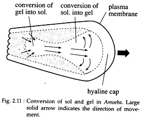
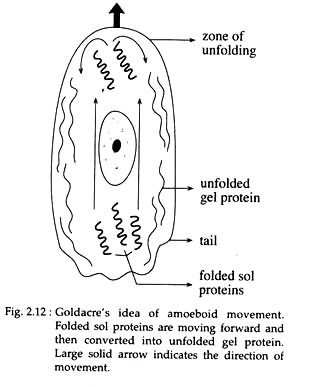
Goldacre and Lorch (1950 & 1952) emphasised that amoeboid movement results from a motive force, exerted by an ATP-mediated contraction of the tail or uroid or the rear part of the cell. This tail contracts as the unfolded gel protein in this region folds into sol protein.
The sol then flows forward and unfolds again at the advancing front region of the cell (Fig. 2.11 & 2.12). A tail organizer — an enzyme, produces the necessary ATP from cytoplasmic precursors.
ii. Frontal Contraction Model:
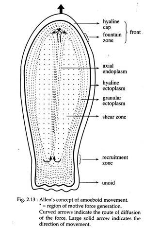
Allen (1961), proposed hat the motive force is generated in the frontal part rather than in the tail of an Amoeba. According to him, a motive pull is generated in a ‘fountain zone’ lying behind the ‘hyaline cap’ at the frontal part of the cell.
The axial endoplasm contracts at the fountain zone and flows posterolaterally below the ectoplasmic tube. It then returns into the axial endoplasm at a recruitment zone, lying just in front of the tail or uroid part of the cell (Fig. 2.13).
The major controversy centres around the question whether the amoeboid cell is pulled along or pushed forward. If the contraction occurs in the ectoplasm of the uroid, then it is argued that the increased pressure on the less structured endoplasm pushes it forward to regions of low pressure.
ADVERTISEMENTS:
If, on contrary, the force-producing contractions occur at the tips of the advancing pseudopods, the viscoelastic endoplasm will be pulled forward into the contraction zone, where endoplasm is converted into ectoplasm.
Either theory requires an explanation of the associated processes by which endoplasm is converted into ectoplasm at the advancing tips of the pseudopods while ectoplasm is converted into endoplasm simultaneously in the uroid.
However, both these concepts have also lost the importance in recent years and amoeboid movement is now known to be the result of sliding interactions between actin and myosin filaments like that of the muscle cells.


