In this article we will discuss about the circulatory system of rattus norvegicus.
Two distinct circulatory fluids, namely blood and lymph, circulate through the circulatory system of the body. Blood flows through well-defined blood vessels and is pumped by an organ called heart. The lymph flows through lymph vessels and intercellular spaces.
Several contractile lymph hearts force the flow of lymph. The blood vascular system is formed by (a) blood, (b) heart, and (c) blood vessels, while the (a) lymph (b) lymph vessels, and (c) lymph hearts constitute the lymphatic system.
Blood Vascular System:
ADVERTISEMENTS:
(i) Blood:
Blood consists of liquid plasma and many corpuscles floating in the plasma. The plasma is a pale yellow fluid and contains various inorganic salts, vitamins and hormones. There are three different types of blood corpuscles. They are erythrocytes, leucocytes and thrombocytes. Erythrocytes or red blood corpuscles or RBC are round and biconcave.
Mature erythrocytes are non-nucleated and contain a red pigment called haemoglobin. Haemoglobin renders red colour to the blood. It has a strong but loose affinity for oxygen. The red blood corpuscles carry oxygen to different parts of the body and carry back carbon dioxide from different parts of the body to the lungs through the heart. Leucocytes or white blood corpuscles or WBC are larger than RBC and their number is much less than RBC.
The leucocytes are of different types and their classification is dependent upon the structure of nucleus and stain-ability of the cytoplasmic granules. These cells move in amoeboid fashion and act as scavengers of the body. Thrombocytes or blood platelets are small and non-nucleated. They occur in groups. In case of injury these cells break down and produce an enzyme which coagulates the blood and thereby prevents loss of blood.
ADVERTISEMENTS:
(ii) Heart:
The heart is located in a space within the thorax between the two pleural bags. The space is known as mediastinum. The heart is covered over by a thin peritoneal membrane called pericardium. Pericardium is two layered. The outer layer is known as fibrous pericardium and the inner layer is called serous pericardium.
The serous pericardium, in reality, is again made up of two layers. The innermost layer adherent to heart is called visceral layer or epicardium and the other (outer) one is called parietal layer. The space between the fibrous layer and serous layer is known as pericardial cavity.
The heart is a muscular, internally hollow, four-chambered and cone-shaped structure that occupies most of the thoracic cavity. It lies roughly in the midline of the cavity with its base located cranially and the apex caudally.
ADVERTISEMENTS:
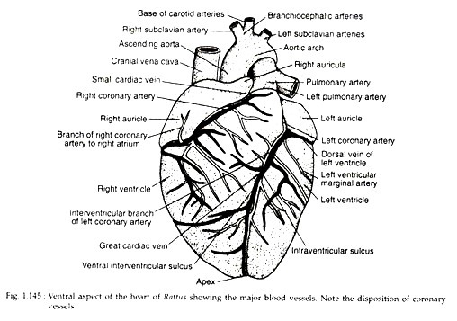
On the surface of the heart there are three grooves or sulci. The coronary sulcus encircles the heart transversely and externally represents the line of separation between the auricles and ventricles. The sulcus is fitted up with coronary vessels and little amount of fat.
The dorsal and ventral inter-ventricular sulci mark externally the line of separation between the two ventricles (Fig. 1.145). There are shallow and small vessels that travel within them.
The heart is four-chambered consisting of two auricles (left and right) and two ventricles (left and right). The right auricle lies cranial to right ventricle and receives the venous systemic blood. An internally located ridge within the right auricle divides it into two regions— sinus venarum cavarnum and right auricula.
The sinus venarum cavarnum is smooth- walled but the auricula is lined by five muscular ridges called pectinate muscles. There are four main openings in the right auricle. Blood enters in the right auricle through three of these openings while through the other, blood passes on to the right ventricle.
The three openings through which blood comes to the right auricle are:
(a) Coronary sinus— It is located between the opening of caudal vena cava and atrioventricular opening.
(b) Larger ostium of caudal vena cava— It is located in the caudal aspect of the auricle near the inter-arterial septum,
(c) Smaller ostium of the cranial vena cava— It is located on the dorsocranial aspect of the auricle.
ADVERTISEMENTS:
Blood from the right auricle flows to the right ventricle through the right atrioventricular ostium. The dorsolateral wall of the right auricle is demarcated by the inter-auricular septum. This septum at its right auricular face bears a small crescent-shaped depression called fossa ovalis. During embryonic stage an aperture called foramen ovale remains present at this spot. This aperture, however, becomes closed before the birth of the animal.
The left auricle is smaller in size than the right auricle and is separated internally from it by the inter-auricular septum. Inside the left auricle, there is the left auricula (auricula sinistra) which is very similar to that of the right auricula. It receives blood through a large pulmonary opening (ostium venerum pulmonalium) located on its dorsal wall. It opens into the left ventricle through the left atrioventricular ostium.
The right ventricle lies caudal to the right auricle. It is thick-walled. It receives blood from the right atrioventricular ostium. The ostium is an oval ring surrounding the right atrioventricular valve. The valve is known as tricuspid valve because it is formed by three delicate and transparent cusps. According to their location the cusps are known as ventral angular cusp, dorsal parietal cusp and medial septal cusp.
The bases of these cusps remain attached to the border of the atrioventricular ostium and their apexes project into the ostium. Each cusp is held in position by chordae tendineae and papillary muscles to prevent back- flow of blood to the right auricle. The chordae tendineae are strong, white and delicate fibres attached to the cusps and anchored to the muscular wall.
The conus arteriosus is fused with the right ventricle and is present as the funnel- shaped cranial portion of the right ventricle leading to the ostium of the pulmonary trunk internally and bordered externally on the right by the right auricula.
The lumen of the ventricle is provided with a number of muscular ridges called trabeculae carnae.
The pulmonary trunk arises from the right ventricle. The round ostium of the pulmonary trunk lies close to the inter-ventricular septum. The ostium is provided with pulmonary valves. Three semilunar transparent cusps constitute the valves. The cusps are known as right, left and intermediate cusps. The valves prevent the backflow of blood to the right ventricle.
The left ventricle is larger than the right ventricle. It is provided with thick walls. The left atrioventricular ostium is provided with bicuspid or mitral valve. This valve is composed of two unequal-sized cusps—a larger septal cusp and a smaller parietal cusp.
The cusps are held in position by chordae that remain anchored to the ventricular wall. The valves prevent the backflow of blood into the left auricle. The lumen of the left ventricle is provided with trabeculae carneae resembling that of the right ventricle.
The left aortic arch arises from the left ventricle. The aortic ostium is the opening of the aorta and it lies near the centre of the base of the heart. The ostium is provided with aortic valves. Three semilunar cusps similar to those of the pulmonary cusps are present. The valves prevent the backflow of blood.
Mechanism of circulation through heart:
The heart works day and night by alternate contraction (systole) and relaxation (diastole). The two auricles begin their systole at the same time and blood from both the auricles is forced into both the ventricles. Deoxygenated blood goes to the right ventricle and oxygenated blood comes to the left ventricle.
The back flow of blood is prevented by the atrioventricular valves. The ventricles thus filled up with blood, start systole. In this phase the atrioventricular valves become closed and blood from the right ventricle is forced through the pulmonary arch while that from the left ventricle is forced through the left aortic arch.
The ventricular systole is followed by a phase of diastole of the whole heart. At this stage the semilunar valves remain closed, deoxygenated blood from the caval veins enters the right auricle and oxygenated blood from pulmonary veins enters the left auricle.
(iii) Blood vessels:
Blood is conveyed from the heart to the different parts of the body through well-developed blood vessels. These vessels form a well-knit circuit inside the body. This type of circulation is known as closed circulation. Oxygenated blood is carried away from the heart by arteries (excepting pulmonary arteries) and deoxygenated blood is carried to the heart by veins (excepting pulmonary veins).
The arteries break up into arterioles which, in turn, break up into arterial capillaries. The arterial capillaries unite with venous capillaries and make up a capillary network. The venous capillaries unite and form venules. The venules form the veins and thus a close circuit is built.
Arterial system:
Two main arches come out from the heart. These are pulmonary and aortic arches. The pulmonary arch or trunk emerges from conus arteriosus of the right ventricle and the left aortic arch emerges from the left ventricle. Semilunar valves are present at the base of each of the openings.
The pulmonary arch carries deoxygenated blood. Soon after its emergence from the right ventricle it bifurcates into right and left branches. The right pulmonary artery passes dorsal to the ascending aorta and then divides into four main branches. Each lobe of the right lung receives one branch. The left pulmonary artery passes ventral to the descending aorta and divides into three branches to enter the three lobes.
Aortic arch:
Only the left aortic arch is present. It originates from the left ventricle. It is the largest artery in the body and the main trunk of the systemic arterial system. For the sake of clarity of description it has been divided into three portions—the ascending aorta, the arch of the aorta and the descending aorta.
The aorta begins as ascending aorta and then curves dorsally and to the left to form the arch or aorta. From the level of second and third thoracic vertebrae it is known as descending aorta. It runs caudally parallel to the vertebral column and bifurcates at the lumbar region of the abdomen forming iliac vessels.
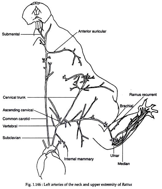
Branches from the different portions of the aortic arch (Fig. 1.146)
A. Ascending aorta:
The ascending aorta gives rise to two or three coronary arteries. The artery that goes to supply the right and dorsal surface of the heart is known as right coronary artery while that which supplies the left side is known as left coronary artery.
B. The arch of the aorta:
From the cranioventral aspect of the arch of aorta arise two large arteries—the brachiocephalic trunk and left subclavian artery (Fig. 1.146). These arteries supply blood to the neck, head and left forelimb. The arch of the aorta is bound to the pulmonary trunk by a fibrous band called ligamentum arteriosum immediately after the origin of the left subclavian artery.
The brachiocephalic artery is rather large and stout. It courses cranially. At the level of the first rib it gives rise to the left common carotid. The brachiocephalic artery continues further cranially and then bifurcates into right common carotid and right subclavian artery.
The two common carotid arteries lie lateral and parallel to the trachea. From both arise similar types of arteries. Both the carotids at the level of the cricoid cartilage of the larynx bifurcate to form the external and internal carotids.
The internal carotids are smaller and they supply blood to the deep structures of the head and the brain while the external carotids supply the respective sides of the head. Of the two subclavian arteries the left one is larger than the right and both course cranio-laterally towards the left and right, respectively.
The subclavian arteries give the following branches:
(a) Costocervical trunk:
It arises from the craniodorsal aspect of the branch and courses dorsally. It divides soon after its origin into 3 branches (cranial) descending scapular, deep cervical, and supreme (caudal) intercostal.
(b) Vertebral artery:
It arises from the craniodorsal border or from the costocervical trunk. It courses cranially for some distance and then curves to take a dorsal position.
(c) Internal thoracic artery:
It originates from the caudoventral border of the subclavian and lies opposite to the costocervical trunk. It courses caudally. The right internal thoracic artery gives rise to percardionephric, bronchial mediastinal and phrenic arteries of both sides. While from the left internal thoracic artery arises only one branch on the left side to the mediastinium.
(d) Superficial cervical trunk:
The superficial cervical trunk is the distal branch of the subclavians. It is a long artery and lies between shoulder and neck.
C. Descending aorta:
The part of the left aortic arch from the level of second and third thoracic vertebrae and the rest is known as descending aorta. It runs caudally parallel to the vertebral column and bifurcates in the lumbar region to form the left and right iliac arteries. For the sake of description it is discussed under two heads—Thoracic subdivision and Abdominal subdivision.
(a) Thoracic subdivision:
The part of the descending aorta lying between second or third thoracic vertebrae and second to fourth lumbar vertebrae is known as thoracic subdivision or thoracic aorta. It runs dorsocaudally along the left of the vertebral column. Eight pairs of dorsal intercostal arteries arise from the dorsal border of the aorta and these extend laterally. Each dorsal intercostal artery sends a dorsal branch and a spinal branch.
The dorsal branch supplies the dorsal and lateral vertebral muscles and the overlying skin while the spinal branch supplies the spinal canal. A number of short twigs called cranial phrenic arteries arise as separate vessels from the aorta near its point of entry into the diaphragm. They supply the dorsolateral peripheral border of the diaphragm.
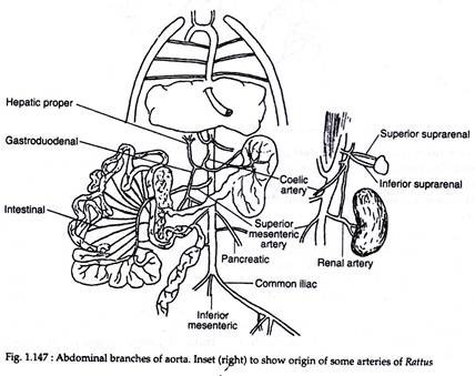
(b) Abdominal subdivision (Fig. 1.147):
The last part of the descending aorta from the level of the diaphragm is known as abdominal subdivision. The branches from the abdominal subdivision (Fig. 1.147) is divided into two categories—paired and unpaired visceral arteries and the paired parietal or lumbar arteries.
I. Unpaired visceral arteries:
1. Coeliac trunk:
The coeliac trunk is the largest artery arising from the abdominal aorta. It is a stout artery. There exists confusion regarding the nomenclature and variability of the different branches that emerge from it.
It gives the following branches:
(i) Gastropancreaticosplenic artery:
It is the first branch of the coeliac artery. Near its origin it gives rise to the left gastric artery which courses cranially and supplies blood to the ventral and dorsal surfaces of the lesser curvature of the stomach. The gastro-pancreatico-splenic artery continues laterally on the craniodorsal side and from it arises the splenic artery. Splenic artery sends many branches (pancreatic branch) to the pancreas.
(ii) Accessory middle coeliac artery:
It arises from a point just distal to the gastropancreaticosplenic artery. It is short and supplies blood to the transverse and proximal colon.
(iii) Hepatic artery:
The branch located caudad to accessory middle coeliac artery is the hepatic artery. From it arises cystic artery that supplies blood to the gall bladder.
(iv) Cranial mesenteric artery:
It is the last and largest branch from the coeliac artery. It supplies blood to most of the intestine and mesenteries.
2. Caudal mesenteric artery:
It is a small branch and arises from the ventral surface of the aorta from a point just caudad to the kidneys. It courses ventrocaudally and supplies blood to the descending colon.
II. Paired visceral branches:
(i) Renal arteries:
There are two pairs of renal arteries—cranial and caudal. They arise from the level of the 2nd and 3rd lumbar vertebrae and supply the cranial and caudal parts of the kidney respectively.
(ii) Testicular artery:
In males, a pair of testicular arteries is present. They course ventrolaterally and supply blood to the male gonads.
In females, these arteries are called ovarian arteries.
(iii) Parietal or Lumbar arteries:
There are six pairs of lumbar arteries that arise from the aorta extending between testicular or ovarian artery and the point of bifurcation of the aorta. They supply blood to the lumbar vertebrae and the body muscles adjoining these vertebrae.
(iv) Common iliac artery:
The abdominal aorta at the level of last lumbar vertebra bifurcates into two and gives rise to common iliac arteries. It diverges caudolaterally from the midline and then divides into two forming internal and external iliac arteries.
From the internal iliac artery arises the prostatic artery in males and vaginal artery in the females. Other branches arising from it supply blood to the reproductive and excretory organs. The external iliac artery enter the thigh part of the hind limb and supplies blood to it.
Venous system:
Deoxygenated blood returns to the heart through veins. Veins follow the same general course as the arteries. Often they bear the same name as the arteries.
The different veins of the body may be grouped in the following way:
(1) Pulmonary group
(2) Cardiac group
(3) Systemic group
(4) Portal group
(1) Pulmonary group:
The pulmonary veins lie ventrocaudal to the pulmonary arteries and dorsal to caudal vena cava. Three groups of pulmonary veins unite to form a single trunk that opens into the left auricle.
The three groups of the pulmonary veins are:
(i) Left cranial group:
It drains blood from the cranial lobe of the left lung.
(ii) Right cranial group:
It drains blood from the cranial lobe of the right lung.
(iii) Common caudal group:
It drains blood from the middle and caudal lobes of the left lung and from the middle and accessory lobe of the right lung.
(2) Cardiac group:
A group of cardiac veins drain blood from the different regions of the heart. They generally follow the coronary arteries and open into the right auricle by coronary sinus or directly by tiny vessels.
(3) Systemic group:
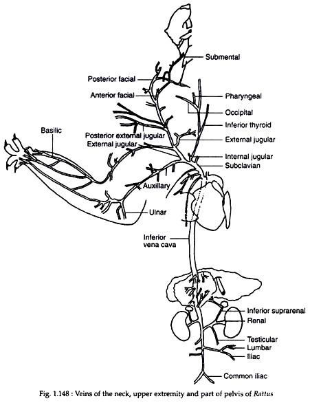
Deoxygenated blood is collected from the anterior and posterior parts of the body and is returned to the right auricle by two large veins (Fig. 1.148). The one that collects blood from the anterior part is known as the cranial vena cava (in old nomenclature it was anterior vena cava or precaval). Blood from the posterior region is brought to the heart by caudal vena cava (posterior vena cava or postcaval in old nomenclature).
Blood from the brain is collected by internal jugular veins (left and right). It comes out of the cranial cavity by the jugular foramen and runs ventrocaudally lateral to the trachea. Each one receives a good number of tributaries from the respective sides and joins with the external jugular vein of the same side at a point where the external jugular vein receives the axillary vein.
The external jugular veins (left and right) originate by the fusion of linguofacial and maxillary veins. It courses downwards and towards the medial border of the first thoracic rib. Here it meets the axillary vein on the lateral side and internal jugular vein on the craniomedial border and forms the brachiocephalic veins. During its downward journey it receives many veins like superficial cervical, prescapular and veins coming from cephalic regions.
There are two brachiocephalic veins— left and right. Each of them has been formed by the union of external and internal jugular veins together with the axillary vein of the corresponding sides. The two brachiocephalics are short and converge at the level of the first rib. Both the branches receive vertebral and costocervical veins. Left supreme intercostal vein opens to the left brachiocephalic and right intercostal to the right brachiocephalic.
The two brachiocephalic veins unite and give rise to the large cranial vena cava. It courses downward and enters the craniodorsal aspect of the right auricle after piercing the pericardium. It receives the common trunk of the internal thoracic vein on the left and the right azygos vein on the right.
The caudal vena cava is formed in the posterior part of the abdominal cavity. The left and right common iliac trunk unite at the level of sixth lumbar vertebra. The common iliac drains the hind limb and the pelvic organs. Each common iliac is formed by the union of internal and external lilacs coming from the hind limb. The internal iliac is short and receives a number of small veins.
The important ones among them are—prostatic vein (in case of males) and vaginal vein (in case of females), visceral and parietal veins, pedal vein, etc. The external iliac vein extends between its point of meeting with the femoral vein within the hind limb and its point of meeting with the internal iliac vein.
It receives a number of small veins. The important ones amongst them are—caudal abdominal vein, deep femoral vein, iliolumbar vein etc. The caudal vena cava runs towards the heart and lies right to the abdominal aorta.
On its journey towards the heart it receives the following paired veins in a postero-anterior sequence:
(i) Six pairs of lumbar veins,
(ii) Testicular vein (in males) or ovarian vein (in females). In some cases these veins open into the renal vein instead of opening into the caudal vena cava,
(iii) Renal vein from the kidneys,
(iv) Cranial abdominal vein,
(v) Suprarenal vein,
(vi) Hepatic vein, and
(vii) Cranial phrenic vein.
(4) Portal group:
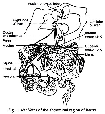
Only hepatic portal vein is present. The hepatic portal vein is formed by the union of a number of small veins coming from the intestine and mesenteries. The important veins that take part in the formation of hepatic portal vein are—lineogastric returning blood from the stomach and spleen, duodenal vein, cranial and caudal mesenteric veins, etc. (Fig. 1.149). The hepatic portal vein enters the liver and breaks into capillaries. Blood is collected inside the liver by hepatic veins which pour their contents in the caudal vena cava.
Lymphatic system:
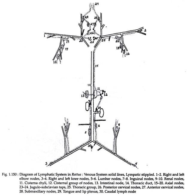
Lymph is a watery liquid containing few leucocytes. It is circulated in the body through lymph vessels. There are a pair of large lymph vessels running one on either side of the dorsal aorta (Fig. 1.150). Small lymph vessels coming from different parts of the body open into these large vessels.
The large vessels in their turn open into the subclavian vein of the corresponding side. Contractile lymph hearts, located on the vessels, help in the propagation of the fluid.
