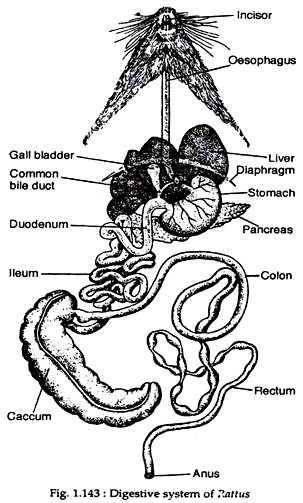
The digestive system of Rattus norvegicus is constituted by the alimentary canal and the digestive glands (Fig. 1.143).
(i) Alimentary Canal:
It is a long tube starting from mouth to anus. But the tube is demarked into different regions. The function of each region is different.
(a) Mouth:
ADVERTISEMENTS:
The alimentary canal begins from the mouth. It is a transverse aperture and is guarded by two soft and movable lips. The upper lip is provided with a cleft in the middle.
(b) Buccal cavity:
ADVERTISEMENTS:
Mouth leads to the buccal cavity. The roof of the buccal cavity is formed by a palate. The anterior portion of the palate is called hard palate. The palate separates the mouth cavity from the nasal passage. The floor of the buccal cavity houses the tongue. The anterior end of the tongue is free and the posterior end is attached with the floor.
The tongue is muscular and movable. The upper surface of the tongue is rough and contains numerous papillae or taste buds. Both the jaws are provided with teeth. The teeth help in ingesting food. Teeth are thecodont, heterodont and diphydont type. Dental formula is 1.0.0.3/1.0.0.3. The incisors are long, chisel-shaped and can be seen from outside.
A tooth has two parts, crown and root. Crown is the visible part and the root remains embedded in a socket on the jaws. The tooth is made up of a substance called dentine. The dentine of the root region remains covered by cement while the crown region of the tooth remains covered by hard and shiny enamel. Each tooth bears an inner pulp cavity. This cavity remains filled up with jelly-like pulp, blood vessels and nerves.
(c) Pharynx:
ADVERTISEMENTS:
Buccal cavity leads to another chamber called pharynx. The dorsal part of the pharynx is called nasopharynx and the ventral part is called buccopharynx. Paired internal nostrils and Eustachian tubes enter into the nasopharynx region. The posterior margin of the soft palate extends into the nasopharynx as velum. The two sides of the velum are with a peculiar lymphoid tissue called tonsil.
A slit called glottis is present on the floor of the buccopharynx just posterior to the tongue. The tongue is a large, elongated muscular organ covering most of the floor of the mouth cavity. The glottis communicates with the respiratory tube and is guarded by a cartilaginous flap called epiglottis. Posteriorly, the buccopharynx opens into the oesophagus through an aperture called gullet.
(d) Oesophagus:
Oesophagus is a long tube running along the mid-ventral line of the neck region. It runs through the thoracic region and after passing through the diaphragm opens into the stomach.
(e) Stomach:
Stomach is a highly muscular and glandular sac (Fig. 1.143). The inner concave side of the stomach is called lesser curvature and the outer convex surface is called greater curvature. The end of the stomach towards the oesophagus is called the cardiac end and its opposite end is called pyloric end.
Due to twisting, the cardiac end has taken a position towards the left and the pyloric end has taken a position towards the right in the abdominal cavity. The opening at the pyloric end is guarded by a valve called pyloric sphincter.
(f) Intestine:
The remaining part of the alimentary canal is known as intestine. It is divisible into duodenum, ileum and large intestine. The duodenum begins from the pyloric end of the stomach and forms a ‘U’-shaped loop. Ileum is much coiled continuation of the duodenum. The coiled loops of the ileum are held in position by folds of mesenteries.
ADVERTISEMENTS:
Ileum opens into the large intestine and the opening is guarded by an ileocoelic valve. The large intestine is wide and is divisible into proximal colon and distal rectum. The colon is coiled and beaded in parts while the rectum is straight. A large blind sac called caecum is present at the point of opening of the ileum into the colon.
(g) Anus:
The terminal part of the alimentary canal is represented by an aperture called anus. The anus is guarded by sphincter muscle.
(ii) Digestive Glands:
The different digestive glands that help the process of digestion are listed below :
(a) Salivary glands:
There are five pairs of salivary glands. Parotid glands are located beneath the cutaneous muscles at the junction of mandible and neck. Mandibular glands are located on the ventral surface of the neck. Major sublingual glands lie ventromedial to the mandibular gland. Minor sublingual gland is located between the last two lower molars and the tongue.
Zygomatic or infra-orbital gland lies in the orbit along the dorso-medial rim of the zygomatic arch. All the glands have separate openings into the buccal cavity through ducts. The secretion of the salivary glands is known as saliva. Saliva helps in moistening food and contains an enzyme called ptyalin.
(b) Liver:
It is a massive gland located beneath the diaphragm and above the stomach. It is a four-lobed structure and remains attached to the diaphragm by falciform ligament. The largest and square lobe of the liver has been termed quadrate lobe. It lies more ventral than other lobes and is divided into two equal sized left and right sub-lobes.
A rectangular left lobe lies dorsal to the quadrate lobe and to the left of the midline. A right lobe which is oval lies to the right of the midline. The smallest of all the lobes is the caudate lobe that lies dorsomedially in association with the lesser curvature and in the angular notch of the stomach.
The secretion of the liver is bile. Bile is kept temporarily stored in a pyriform gall bladder lodged in the quadrate lobe of the liver. A common bile duct formed by the union of hepatic duct (from liver) and cystic duct (from gall bladder) carries bile to the duodenum.
(c) Pancreas:
It is a whitish, elongated and irregular-shaped gland located between the limbs of the duodenum. The secretion of the pancreas is known as pancreatic juice. The juice is carried to the distal part of the duodenum by a pancreatic duct.
(d) Gastric glands:
Innumerable gastric glands are present along the inner lining of the stomach. The juice produced by these glands is known as gastric juice.
(e) Intestinal glands:
Numerous glands are present in the inner lining of the duodenum and intestine.
(f) Spleen:
It is an elongated brown coloured organ. It is morphologically connected with the alimentary canal, being situated on the dorsal side of stomach by a fold of mesentery. Spleen is devoid of any duct, and does not produce any hormone. It is believed that it destroys old red blood corpuscles.
