In this article we will discuss about the male and female reproductive organs of Rattus Norvegicus.
(A) Male Reproductive Organs:
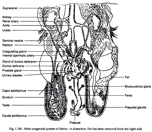
The male reproductive organs (Fig. 1.156) are Testes, Epididymis, Deferent ducts, Spermatic cords, Urethra, Penis, Accessory genital glands, Perineal glandular modifications and Caudal glands.
(i) Testis:
ADVERTISEMENTS:
In the young males the testes remain inside the abdomen. In adults, the testes descend down and remain lodged in a special fold of skin called scrotum. The scrotal cavity and the abdominal cavity remain in communication with each other through a canal called inguinal canal. The paired testes lie in the scrotal pouch and produce sperms.
Each testis is oval in outline and its long axis remains oriented craniocaudally. The testis remains suspended within the pouch by mesorchium (a fold of peritoneum) dorsally and caudally by a fibrous ligament called gubernaculum. Each testis is divided internally into lobules by many thin septa. Each lobule contains tiny seminiferous tubules.
(ii) Epididymis:
ADVERTISEMENTS:
Each testis is associated with an epididymis. The epididymis is a highly convoluted tube consisting of a head, a body and a tail. Head is the largest and most cranial portion of it. The body is narrow and runs along the medial border of the testis. The caudal part opens in the ductus deferentus. The function of the epididymis is to store sperms for ejaculation. Some workers are of the opinion that the epididymis helps in the maturation of sperms.
(iii) Ductus deferens:
The paired deferent ducts are 40 to 60 mm long and are divisible into a coiled epididymal portion and an uncoiled portion. They open separately in the urethra. The openings are located ventral to seminal vesicles, dorsal to the neck of the bladder and medial to the ducts of the coagulating and prostate glands.
The two ducts then open into a median slit within the urethra. Each deferent duct is supported by a mesoductus deferens which extends through the spermatic cord as a separate fold of peritoneum.
ADVERTISEMENTS:
(iv) Spermatic cord:
The two spermatic cords (one each for the deferent ducts) contain the deferent ducts and their mesenteries. Each spermatic cord begins at the inguinal ring and ends within the scrotal pouch.
(v) Accessory glands:
The accessory genital glands consist of the following—seminal vesicles, the coagulating glands, the ventral and dorsal lobes of the prostate gland and bulbourethral gland. A common sheath envelops the coagulating glands, the prostate and the urethral end of the different ducts and seminal vesicle.
(a) Seminal vesicle:
Paired seminal vesicles are the largest amongst the accessory glands. Each seminal vesicle is a cylindrical and elongated structure the free end of which may be bifid. They converge medially and enter the urethra by a pair of ducts that lie dorso-caudal to the deferent ducts. The lumen of the seminal vesicle remains filled up with a milky white fluid and its walls are granular.
(b) Coagulation glands:
These are paired and pyramid-shaped bodies. They lie latero-dorsal dorsal and in close proximity to the seminal vesicle. Each gland has a single duct that opens into the urethra craniolateral to the opening of deferent ducts and seminal vesicles. The secretion of these glands coagulates the secretion of the seminal vesicle and produces vaginal plug.
ADVERTISEMENTS:
(c) Prostate gland:
It consists of two pairs of lobes—large dorsal and small ventral and the lobes are joined by isthmus. It lies caudomedial to the coagulation gland. A single pair of ducts comes from the ventral lobe and many pairs of ducts come from the dorsal lobe. These ducts open into the urethra and the openings are located craniolateral to the opening of the coagulating gland.
(d) Bulbourethral glands:
These paired glands are small, lobulated and lie ventrolateral to the rectum. From each gland a duct arises and the ducts open into the urethra on the dorsal surface. Besides these glands a peculiar structure called uterus masculines is present in male guinea-pig. It is considered homologous to the uterus of the female. This is a flat bilobed, hollow organ that lies between the mesentery connecting the deferent ducts and that of the two seminal vesicles and its unpaired central body opens into the urethra.
(vi) Urethra:
The male urethra differs functionally from the female urethra because it transports seminal fluids as well as urine. The urethra is divided into two portions— pelvic portion and spongy portion. The pelvic portion extends from the neck of the urinary bladder to the penis and receives most of the ducts of accessory glands.
The spongy portion remains embedded within the corpus spongiosum of the penis. It opens externally through the urethral orifice located at the tip of the penis.
(vii) Penis:
Penis is the male copulatory organ. It is eversible but during sexual inactivity lies retracted within a sheath of skin called preputial sheath. Penis is made up of two parts—body and glans. The body is composed of two layers of corpora cavernosa on the dorsal surface and its ventral midline is made up of corpus spongiosum which houses the spongy portion of the urethra.
The penis is enveloped by tunica albugenia. The glans are shorter than the body. It is cylindrical and ends in a rounded tip. The tip bears the urethral orifice. The os penis or baculum is present on the dorsal surface of the glans.
(B) Female Reproductive Organs:
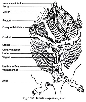
The female reproductive organs (Fig. 1.157) consists of a pair of ovaries which produce ova or eggs; two oviducts which carry the ova to the uterus; a uterus which supports the developing guinea-pigs, and a vagina which is a passage between uterus and the external genitalia or the vulva. The mammary glands may be considered under the system as they are functionally associated with these organs.
(i) Ovaries:
There are two ovaries. They are intra-peritoneal, oval and dorsoventrally flattened. The right ovary is caudolateral to the right kidney and the left ovary is cranio-lateral to the left kidney. The long axis of the ovary lies parallel to the axis of the animal. Each ovary has a medial and a lateral surface and a cranial tubal and caudal uterine extremity. The straight medial border and the tubal extremity is invested by mesovarium.
(ii) Oviducts:
The oviducts or Fallopian tubes are paired and lie along the lateral regions of the ovary. Each oviduct consists of three parts— the infundibulum, the ostium and the tubal part. The infundibulum is the most cranial part of the oviduct. It is triangular, flared and its border is provided with many papillary elevations called fimbrae.
The ostium is the small opening present in the oviduct at the cranial apex of the infundibulum. The tubal portion is a thin coiled tube; the terminal part of it joins with the uterine horn.
(iii) Uterus:
Uterus is bicornuate and is the largest organ of the female reproductive tract. It is a ‘Y’-shaped structure extending between the oviducts cranially and vagina caudally. It has two horns, a body and a cervix. The cervix opens into the vagina.
(iv) Vagina:
It is a pink-red canal extending between cervix and vulva. The vagina is dorsoventrally flattened and bears on its internal side a number of longitudinal ridges.
(v) Vulva:
It is the external female genitalia and includes clitoris, the vaginal orifice and labia. The clitoris is homologous to the male penis. It consists of paired roots, body and glans. The vaginal orifice is ‘U’-shaped and usually remains closed by a vaginal closure membrane. The labia are a pair of lateral thick folds of skin bordering the vaginal orifice.
(vi) Mammary glands:
A single pair of mammary glands are located on either side of the midline of the caudal abdomen near inguinal region. During pregnancy the glands become large and start producing milk following parturition. The glands are apocrine in nature. In males they remain rudimentary and non-functional.

