The following points highlight the top two types of limb bones. The limb bones are: 1. Fore Limb 2. Hind Limb.
Type # 1. Fore Limb:
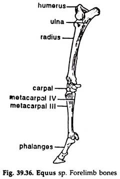
1. The bones of the forelimb consist of a humerus, a radius and an ulna, carpal and metacarpal bones and phalanges (Fig. 39.36).
2. Humerus:
It has a short, rounded and slightly bent shaft. The proximal end bears a tuberosity and a head to fit into the glenoid cavity. The head is separated from the tuberosity by a bicipital groove. The distal end bears a pulley-like structure and a depression just anterior to it.
ADVERTISEMENTS:
3. Radius and ulna:
It consists of two narrow, elongated, separate bones—the radius and the ulna. The radius is slender and smaller .than the ulna and bears a concave facet at the proximal end. The distal end is almost flat. The ulna is long and bears an olecranon process and a sigmoid notch for the humerus.
4. Carpal bones:
These are seven bones of the wrist arranged in two rows. The proximal row contains pisiform, cuneiform, lunar and scaphoid; and the distal row contains trapezoid, magnum and uniform.
ADVERTISEMENTS:
5. Metacarpal bones:
These are the bones of the palm. The first and fifth are completely degenerated. The 2nd and 4th are represented by narrow splint bones. The third one is a large, elongated, round bone, known as cannon bone.
6. Phalanges:
These are the bones of the fingers. The 2nd and 4th fingers are represented by small bones, the sesamoids. The third finger is elongated and consists of three phalanges, the last one bearing a hoof.
Type # 2. Hind Limb:
1. The bones of the hind limb consist of a femur, a tibia and fibula, tarsal and metatarsal bones and phalanges.
2. Femur:
It has a long, stout, slightly curved shaft. The proximal end bears a prominent head to fit into the acetabulum of the pelvic girdle. Three elevated areas— greater trochanter immediately below the head and lesser and third trochanter below the greater trochanter, are present. The distal end is pulley-like. The condyles are separated by an inter-condylar notch. A patella is present at the knee joint.
3. Tibia and fibula:
It consists of two separate bones attached at the ends only. The tibia is strong, stouter at the anterior end and narrower towards the posterior end. Two concave facets are present at the proximal end. A cnemial crest is present towards the proximal end. The distal end also bears concavity. The fibula is slender and narrows towards the distal end.
4. Tarsal bones:
These are the bones of the ankle and are six in number. The astragalus has a pulley-like surface above for articulation with the tibia. Its distal surface is flattened and articulates more with the navicular than with the cuboid. The calcaneum does not articulate with the fibula. The fused meso-, ento- and ecto-cuneiform articulate distally with the metatarsals.
5. Metatarsal bones:
These are the bones of the foot. The first and fifth are completely degenerated. The second and fourth are represented by narrow splint bones. The third one is a large, elongated, round bone known as cannon bone.
ADVERTISEMENTS:
6. Phalanges:
These are the bones of the fingers. The 2nd and 4th fingers are represented by small bones, the sesamoids. The third finger is elongated and consists of three phalanges, the last one bearing a hoof.
