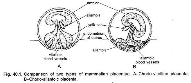
The chorio-allantoic placenta has been classified into various types on the basis of its morphology, arrangement of villi, histology and electron microscopy.
A. Morphological Classification of Placenta:
On the basis of closeness of foetal and maternal tissues, placentae may be of the following three types:
1. Non-Deciduous Placenta or Semiplacenta:
In most mammals, the implantation is superficial, i.e, the blastocyst lies in the cavity of the uterus in contact with the uterine wall. The contact may be made more intimate by the surface of the blastocyst by forming finger-like outgrowths which penetrate into depressions in the wall of the uterus. Such outgrowths are initially formed by the trophoblast (i.e., the epithelial layer covering the blastocyst), but later on the connective tissue and blood vessels invade the outgrowths.
ADVERTISEMENTS:
These outgrowths are called chorionic villi, the blood vessels of chorionic villi are the branches of allantoic blood vessels in case of chorio-allantoic placenta. (In chorio-vitelline placenta, vitelline blood vessels give their branches to chorionic villi).
At the time of birth, when parturition (the separation of the foetus and its membranes from the mother’s body) occurs, the chorionic villi are simply drawn out from the depressions in the wall of the uterus and, thus, maternal and foetal tissues are separated without further damage to the uterine wall and no bleeding occurs.
This type of placenta is called non-deciduate or non- deciduous placenta and is found in pigs, cattle and some other mammals. Further, the chorionic villi of a non-deciduate placenta, because lie in apposition with the endometrium, but, do not fuse with it, so such a placenta is also called semiplacenta.
2. Deciduous Placenta or Placenta Vera:
ADVERTISEMENTS:
In other mammals, however, the degree of intimacy between maternal and foetal tissues becomes further increased. The wall of the uterus becomes eroded to various degrees through the action of the trophoblast and the embryonic tissues penetrate into the uterine wall, establishing a more intimate contact and facilitating the passage of substances from the mother to the foetus and from the foetus to the mother.
Here because the chorionic villi fuse with the eroded uterine mucosa, such placenta is called placenta vera (true placenta). At the end of pregnancy the uterine wall is no longer intact and when the foetus with its membranes including the chorion is removed, more or less extensive haemorrhage from the uterine wall ensues (i.e., at birth, when such placenta is discharged, the uterine lining also tears away with some bleeding). Such a type of placenta found in higher eutherian mammals and is called deciduate or deciduous placenta.
The maternal tissues which are expelled at birth in the case of deciduate placenta are called deciduae. The haemorrhage at parturition is normally stopped by the same mechanism as serves for the expulsion of the newborn, the contraction of the muscular wall of the uterus constricts the blood vessels and, thus, slows down the flow of blood, until clotting of the blood stops the haemorrhage altogether.
ADVERTISEMENTS:
3. Contra-Deciduate Placenta:
In Perameles and Talpa (mole), somewhat modified type of deciduate placenta occurs, which is called contra-deciduate placenta. In such case, not only there is a loss of maternal tissue but also of the foetal portion of the placenta, both of which absorbed in situ by maternal leucocytes.
B. Classification of Placentae According to the Distribution of Villi on Chorion:
In different mammals the pattern of distribution of villi varies from species to species and accordingly following kinds of placentae have been recognised:
1. Diffuse Placenta:
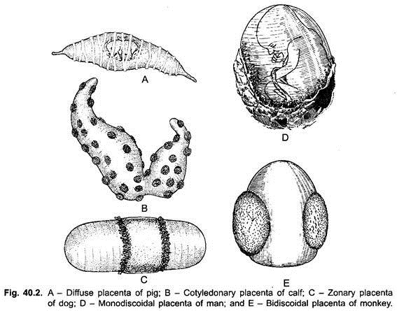
In some mammals (e.g., ungulates, pig, sow, mare, horse, lemur, etc.) the chorionic villi remain scattered all over the surface of the chorion and their placentae are correspondingly expensive. Such placentae are called diffuse placentae.
2. Cotyledonary Placenta:
In a cotyledonary placenta, the villi are found in groups or patches, while the rest of the chorion surface is smooth. The rosettes or patches of villi are called cotyledons, and the placenta of this type is found in ruminants (cud-chewing), ungulates such as cattle, sheep and deer. Camel and giraffe have an intermediate type of placenta in which villi are arranged in cotyledons as well as scattered.
3. Zonary Placenta:
ADVERTISEMENTS:
In a zonary placenta, the villi are developed in the form of a belt or girdle-like band around the middle of their blastocyst or chorionic sac, which is more or less elliptical in shape. Such a placenta occurs in carnivores (e.g., cats, dogs, etc.). Raccoon has incomplete zonary placenta.
4. Discoidal Placenta:
In insectivores, bats, rodents (mouse, rat, rabbit, etc.) and bear, the villi are restricted to a circular disc or plate on the dorsal surface of blastocyst.
5. Metadiscoidal Placenta:
In primates also discoidal placenta is found but of special type, i.e., chorionic villi are at first scattered but later on become restricted to one or two discs. Thus, in man the placenta has a single disc-shaped villous area and is called monodiscoidal placenta. In the monkeys, the placenta consists of two disc-shaped villous areas and such a placenta is called bidiscoidal placenta.
C. Classification of Placenta According to Histology:
On histological basis, following types of mammalian placentae have been recognised:
1. Epithelio-Chorial Placenta:
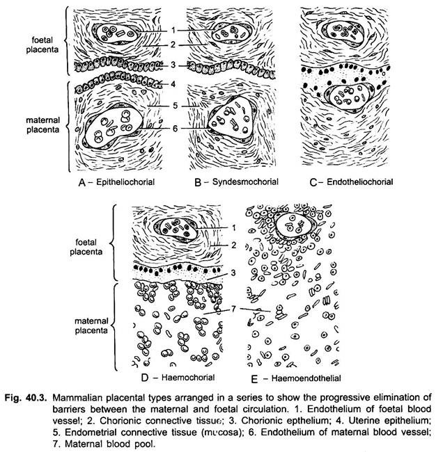
The epithelio-chorial type placenta is most primitive type and it is found in marsupials, ungulates (pig, horse, cow, cattle, etc.) and lemurs.
In this case, placenta is formed of six tissue or membranes:
(i) The endothelium of the maternal blood vessels;
(ii) Endometrial connective tissue (mesenchyme);
(iii) Uterine epithelium;
(iv) The ectoderm of the chorion or chorionic epithelium;
(v) Chorionic connective tissue (foetal mesenchyme) and
(vi) The endothelium of foetal blood vessels.
Because, the immediate contact of the two halves of the placenta involves chorionic epithelium and uterine epithelium, this type of placenta is called epithelio-chorial placenta. The villi of an epithelio-chorial placenta, push in the wall of uterus and lie in pocket-like depressions of the uterine wall.
2. Syndesmo-Chorial Placenta:
In the ruminant ungulates (cattle, sheep), the foetal and maternal components are fused so intimately as to result in a destruction of the uterine epithelium, thus, bringing the chorion into contact with the connective tissue of the uterine mucosa. Only five barriers or tissues, therefore, lie between the two (viz., foetal and uterine) blood streams. This type of placenta is called syndesmo-chorial placenta.
3. Endothelio-Chorial Placenta:
In carnivores (dogs, cats, bears, etc.), the uterine mucosa is also reduced and the chorionic epithelium comes in contact with endoethelial walls of the maternal (uterine) blood vessels. In such a case, therefore, there lies only four barriers between the foetal and maternal blood streams. This type of placenta is called endothelio-chorial placenta.
4. Haemo-Chorial Placenta:
In the haemo-chorial placenta of primates, insectivores (moles, shrews), and chiropterans (bats), a reduction of the barriers to three occurs, i.e., the endothelial walls of maternal (uterine) blood vessels also disappear and the chorionic epithelium is bathed directly in maternal blood sinuses. Actually, the chorionic villi are surrounded by spaces (sinuses) devoid of endothelial lining, into which maternal blood enters through the uterine arteries flows out through the uterine veins.
5. Haemo-Endothelial Placenta:
In haemo-endothelial placenta of higher rodents (rat, guinea pig, rabbit), the number of barriers between the maternal and foetal blood streams is further reduced to two. In them, the chorionic villi lose their epithelial and connective tissue layers to such a degree that, in most places, the bare endothelial lining of their blood vessels alone separates the foetal blood from the maternal blood sinuses.
D. Classification of Placentae According to the Mode of Implantation:
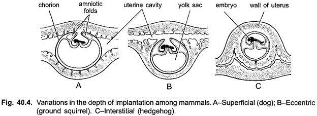
The relation of the chorionic sac to the uterine wall varies greatly among placental mammals.
In general, following three types of implantation may be distinguished, although transitional conditions occur:
1. Superficial Implantation:
Growth of the chorionic sac brings it into contact with the lining of the main uterine cavity. This type of implantation is called central implantation, e.g., ungulates, carnivores, monkey.
2. Eccentric Implantation:
The chorionic sac lies for a time in a fold or pocket which looses off from the main cavity, e.g., beaver, rat, squirrel.
3. Interstitial Implantation:
The chorionic sac penetrates into the substance of the uterine lining, e.g., hedgehog, guinea pig, some bats, ape and man.


