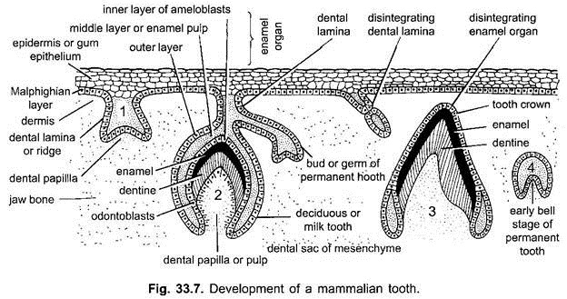
In mammals, teeth develop in the gum or the soft tissue, covering the borders of premaxillae, maxillae and denteries (Fig. 33.7). As in all vertebrates, development of a tooth is similar to that of a placoid scale. Enamel of tooth is derived from epidermis, while the rest of tooth front dermis or mesenchyme.
In the beginning of development of a tooth, there is a thickening of ectoderm along the margin of jaw bone. The basal layer of ectoderm, the Malpighian layer, forms a continuous ridge-like vertical invagination into the underlying dermis. This forms the dental lamina which retains its connection with the outer epidermis.
Mesodermal cells multiply rapidly beneath the ectodermal in growth or dental lamina forming a series of solid bud-like outgrowths at intervals, called tooth germs. The number of tooth germs is as many as the number of milk teeth. In each tooth germ, the inverted cup-like epithelial cap will secrete the enamel hence, termed the enamel organ. This gives rise to the enamel of the tooth.
The outer columnar cells of enamel organ become differentiated into odontoblasts which secrete a layer of dentine on their outer surface. The cells of inner epithelial layer of enamel organ similarly become ameloblasts which form a cap of hard enamel around the top and sides of dentine. No enamel is deposited in the root. Dental papilla is retained as pulp. The central cavity of pulp goes on increasing to become pulp cavity.
ADVERTISEMENTS:
The nerves and blood vessels enter the pulp cavity through the basal opening. Up to this stage the tooth remains inside the tissue (gum). In the later stage its eruption (the eruption of dental papilla) through the overlying epidermis is known as cutting of tooth. Around the root of tooth appears cement or cementum which is a modified bone. Odontoblasts become inactive when tooth is fully formed. However, in rodents, lagomorphs, etc., the odontoblasts remain active throughout life and teeth continue to grow.
