The following points highlight the six main stages involved in interaction between any parasite and its host. The stages are: 1. Host-Symbiont Contact 2. Establishment of the Symbiont 3. Escape from the Host.
Stage # 1. Host-Symbiont Contact:
To be a parasite, an organism must make contact with its host, either actively or passively at a particular stage of its life cycle. Sometimes, the infective form of the parasite approaches the host and makes contact or sometimes the host makes contact.
In the first instance, the active contact may be:
(i) Accidental, e.g., the settling of hydroids on the shell of marine bivalves;
ADVERTISEMENTS:
(ii) Influenced by chemo-taxis, e.g., different amino acids, salts, sugar galactose, bytiric acid etc. act as chemo-attractant,
(iii) Influenced by other taxes, e.g., negative geotaxis and positive photo-taxis of miracidia bring them to their molluscan host; or
(iv) Influenced by visual or chemo-sensitivity of the parasites.
If the infective organism is passive, in majority of instances its mode of contacting the host is dependent on the latter’s feeding habit. For example, human get infected with Entamoeba histolytica when its non-motile cyst is ingested as a contaminant of food or drink.
ADVERTISEMENTS:
Once the contact is made, some endoparasites have to take preparation for entry into host’s particular organ. For example, shedding of ciliated epidermis is found in miracidian larvae of some trematodes for successful penetration into the snail (intermediate host).
Many parasites that gain entry by penetrating the surface of their host, perform lytic secretion that alter hosts’ tissue in a number of ways. Schistosoma mansoni cercariaee secrete hyaluronidase and collagenase like enzymes to dissolve the intracellular cementing substances, thus rendering the intercellular spaces amenable to passage.
Stage # 2. Establishment of the Symbiont:
Even if a parasite makes contact with a potential host, there is no assurance that it will become established and continue normal growth and development. A number of factors influence successful establishment of the parasite within the host.
(a) Habitat selection and attachment:
ADVERTISEMENTS:
Both ecto- and endoparasites are very specific in their habitat selection. Adult Fasciola spp, usually occur in the bile ducts of their definitive host. Likewise, some lice are found only at certain sites on host’s body. Habitat selection is based on biochemical, physiological and physical needs.
In addition to occupying a compatible site, each parasite must become mechanically attached. Attachment not only must prevent involuntary shifting of the parasite from its habitat, but in some cases is essential for its physiologic well- being. Such as, the tapeworm Echinococcus granulosus become embedded in its definitive host’s mucosal crypts with the scolex.
(b) Overcoming the host’s defence mechanism:
The most important factors behind the successful establishment of parasites, especially endoparasites, is their ability to overcome the host’s internal defence mechanism. Successful parasites display an astonishing array of tactics and evade protective immunity by reducing their immunogenicity and inhibiting host immune responses.
Here some examples of parasitic evasion are cited:
i. The particular location of the parasite within the host’s body may provide some protection against host defences. The lumen of intestine is one such site. Among different immunoglobulines, IgA is secreted into intestinal lumen which is not a potent effector molecule against worms that inhabit there. Besides, complement and phagocytic cells are not found in intestine, thus some helminthic parasites are sheltered from cell-mediated immune mechanism of the host.
ii. Protozoan parasites may conceal themselves from the immune system by living inside the host cells or by developing cysts that are resistant to immune effectors of the host.
iii. Schistosoma larvae develop a tegument while travelling to lungs of infected animals that is resistant to damage by complement and cytotoxic T lymphocytes of the host.
iv. Parasites may shed their antigenic coats, either spontaneously or after binding with specific host antibodies. It renders the parasite’s resistant to immune-effector mechanisms.
ADVERTISEMENTS:
v. Parasites may change their surface antigens during their life cycle in vertebrate hosts. In one case, the mature tissue stages of parasites produce different antigens than the infective stages do.
For example, the infective sporozoite stage of malarial parasites is antigenically distinct from the merozoites that reside in the host tissue causing chronic infection. When the host’s immune system responds to infection by sporozoites, the parasite differentiates and expresses new antigens which are no longer a target for immune elimination.
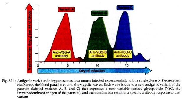
Continuous variation in major surface antigens is also shown by African trypanosomes. In such case, infected individuals show waves of blood parasitemia, each wave consists of parasites expressing a surface antigen that is different from the previous wave (Fig. 6.14).
Thus, by the time the host produces antibodies against the parasites, an antigenically new organism has grown out. Such continuous antigenic variations make it difficult to effectively vaccinate the infected individuals.
vi. Some parasites may absorb host’s antigen, e.g., blood fluke Schistosoma absorbs many host antigens so that the host immune system ‘sees’ only self, not recognising the parasite as foreign.
(c) Deriving adequate nourishment:
Most endoparasites live in an environment where free oxygen is lacking or at a minimum. The utilisation of lipids and proteins as fuel for energy production via the citric acid cycle requires oxygen. Therefore, such parasites depend, at least in part, on glycolysis and fermentation, both being anaerobic processes, for the production of energy.
A number of tapeworms and trematodes are known to feed on partially digested foods of their hosts as well as on mucus and mucosal cells derived from host’s intestine. Thus, these parasites are nutritionally dependent upon their host’s digestive enzymes.
Many intracellular protozoa lack mitochondria in their cytoplasm. Under electron microscope, host mitochondria can be seen clustered around each parasite, indicating that these parasites are dependent upon the mitochondrial respiratory activities of their host for energy production.
(d) Pathogenic changes induced by parasites upon hosts:
The parasites always try to improve their survival value and perpetuate their own species. Therefore, it will be deleterious to a parasite to cause the death of its host, at least prior to its escape or that of its germ cell-bearing progeny.
Nevertheless, many parasites do cause some degree of alteration within their hosts, some of which may result is diseases. Actually the onset of recognisable disease symptoms is dependent on the presence of a sufficient number of parasites as well as physiological conditions of the host.
Here some recognisable changes in host body developed as a result of parasitic interaction are being discussed:
i. Tissue damage:
Some parasites injure the host’s tissues during their entry, e.g., the infective larvae of hookworm inflict extensive damages to cells and underlying connective tissues during penetration of the host’s skin.
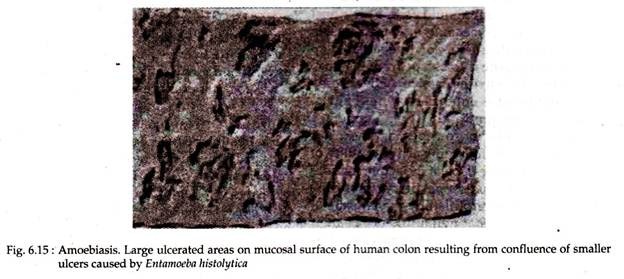
Some inflict tissue damage following their entry, e.g., the amoebic dysentery-causing protozoa, Entamoeba histolytica actively lyses the epithelial cells lining the host’s large intestine causing ulceration that also invites bacterial infection (Fig. 6.15); still others induce histopathology changes by eliciting cellular immunologic response to their presence.
Larvae of Ascaris lumbricoides when pass through lungs of host, cause tissue damage or induce a profound immunologic reaction of asthmatic nature.
Mechanical interference by parasites may in some cases result in injuries to hosts. In human infected with the filarial nematode, Wuchereria bancrofti, the adult worms are lodged in the lymphatic ducts.
The continuous increase in the number of the worms, coupled with the aggregation of connective tissue may result in complete blockage of lymph flow; excess fluid behind the blockage then seeps through the walls of lymph ducts into the surrounding tissues, causing edema of limbs, breasts or scrotum.
ii. Tissue change:
Parasites may bring several types of changes in associated host tissues, such as:
Hyperplasia:
An accelerated rate in division, resulting from an increased level of cell metabolism, is called hyperplasia. The liver fluke Fasciola spp may cause thickening of bile ducts.
Hypertrophy:
Intracellular parasites may cause hypertrophy in cell size. The erythrocytic phase of Plasmodium spp may cause hypertrophy of red blood cells.
Metaplasia:
Changes of one type of tissue into another without the intervention of embryonic tissue are called as metaplasia. The liver fluke Paragonimus sp, when occur in human lungs, becomes surrounded by a wall of host tissue composed of epithelial and fibroblast cells that are not normal constituents of human lungs.
Neoplasia:
When growth of cells in tissue forms a new structure, i.e., tumor it is referred to as neoplasia. Several species of parasites are known to produce tumor in human body, like Schistosoma mansoni can cause tumors in liver and intestine, S. haematobium in bladder and Echinococcus granulosus produce tumor in human lungs.
iii. Effects of toxin/poison:
Many toxic or poisonous substances are secreted, egested or excreted by the parasites that may cause severe damage to hosts. The nematode Parascaris secrete some toxins that irritate the host’s cornea and mucous membranes of nasopharyngeal cavity. Likewise, pathogenic Entamoeba, releases certain potent toxins that cause damage to parasitized host.
iv. Induction of growth:
Parasites are generally considered to be detrimental to their hosts and to cause loss of energy and poor health. Evidences of reverse phenomena are known to happen both vertebrate and invertebrate hosts.
Rats parasitized with larvae of tapeworm Spirometra and blood protozoa Trypanosoma lewisi, respectively, grow faster than the non-parasitized one. The parasites produce a growth-hormones like substance in the former case. Similarly, larvae of the beetle, Tribolium, attain larger sizes when parasitized by the protozoan, Nosema.
v. Sex reversal:
Parasites may cause changes in Sex related characters – a phenomenon called sex reversal. It results mainly by the destruction of gonadal tissues (secondary sex organs) by the parasite. These activities are known as parasitic castration. Sex reversal is common in invertebrates, e.g., male crabs parasitized by the crustacean.
Sacculina, acquire secondary female characteristics like broader abdomen, smaller chelate and egg grasping appendages. On the other hand, larvae of trematode, Zoogonus lasius may cause total loss of gonadal cells in the mudflat snail, Ilyanassa obsoleta. These larvae, also known as sporocysts, do not possess mouth and thus destroy the gonadal tissue by chemical means resulting in direct chemical castration.
Stage # 3. Escape from the Host:
From the view-point of perpetuation of the species and symbiosis, it is a must that the symbiont or parasite should escape their host as it is to make contact and/or enter. Thus, in the host-parasite interaction, the last role on the part of parasite is to escape from the host.
Escape may be active or passive. Active escape of the symbiont is enhanced by mechanical pressure on the part of the host. Temporary ectoparasites like mosquitoes and acarines escape their hosts following active motility of their host. Passive escape of the symbiont does not involve any active participation of the host.
It is best exemplified by the passage of the eggs of intestinal helminths out of their hosts in faeces. However, in some cases, this may involve activity on the part of the parasites. For example, although the eggs of human pin worm are passed out through the host’s anus, the female worms migrate posteriorly to deposit their eggs in the perianal zone.
In addition to active and passive modes of escape, many characteristic patterns of symbiotic escape are found in parasite life. The fertilised eggs of the clams, belonging to the genus Lamsilis are maintained within a specialised gill chamber of the parent known as marsupium or brood chamber. In these chambers, the eggs develop into glochidia larvae.
These parasitic larvae are released from their parent in bursts and become attached to the gills of their host fishes where they feed on superficial tissues and blood. After a period of parasitic existence, the glochidia develop into young adults, drop off and mature into free living adults. In this way Lamsilis clams exhibit a spectacular escape mechanism.
From the above discussion it appeared that to become successfully established in host, the parasites have to undergo all or some stages of interactions. Otherwise, the host simply would not be susceptible and no parasitism or parasitic relations would be established between the symbionts.

