In this article we will discuss about:- 1. Description of Neanthes 2. Locomotion in Neanthes 3. Digestive System 4. Feeding 5. Digestion and Absorption 6. Blood Vascular System 7. Excretory System 8. Nervous System 9. Reproductive System 10. Development 11. Metamorphosis.
Contents:
- Description of Neanthes
- Locomotion in Neanthes
- Digestive System of Neanthes
- Feeding in Neanthes
- Digestion and Absorption in Neanthes
- Blood Vascular System of Neanthes
- Excretory System of Neanthes
- Nervous System of Neanthes
- Reproductive System of Neanthes
- Development of Neanthes
- Metamorphosis in Neanthes
1. Description of Neanthes:
Neanthes (previous name Nereis) belongs to order Polychaeta, class Chaetopoda, phylum Annelida. It is commonly known as sandworm because it is found buried in sand, and also clam worm, as it lives with clams. It is cosmopolitan in distribution.
ADVERTISEMENTS:
Neanthes lives in shallow seas, in rock crevices or hidden under the stones or sea weeds. Some live in tubular burrows of loose tubes made up of mucus in sand or mud at tide level. It is a carnivore, feeding on dead fishes, molluscs, etc.
It is active during night and remains passive in daytime.
2. Locomotion in Neanthes:
ADVERTISEMENTS:
1. Locomotion in Neanthes is brought about by the combined action of musculature, Para podia and the hydrostatic action of the coelomic fluid.
2. Neanthes shows creeping and crawling movements but sometimes it swims.
3. The creeping movements are carried out by the action of Para podia only:
i. Each Para podium performs two types of strokes,—back stroke or effective stroke and recovery or forward stroke.
ADVERTISEMENTS:
ii. In the effective or back stroke the aciculum of Para podium is extended and lowered to come in contact with the substratum and moved backwards against the substratum.
iii. In the recovery stroke, the aciculum is retracted, and the Para podium is lifted above and moved forward.
iv. The combined effective and recovery strokes of numerous Para podia propel the worm forward.
v. The Para podia of two sides work alternately causing successive waves along each side of the worm.
4. The undulatory movement of the body musculature also helps the worm to crawl or swim rapidly:
i. The waves of contractions of the longitudinal muscles of the body wall cause undulation of the body.
ii. The longitudinal muscles of one side contract when the Para podia of that side are moved and the muscles relax when Para podia sweep backwards, which makes crawling effective.
3. Digestive System of Neanthes:
The digestive system consists of an alimentary canal and oesophageal, gastric and intestinal glands.
ADVERTISEMENTS:
Alimentary Canal:
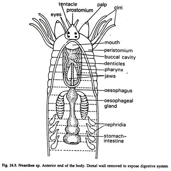
The alimentary canal (Fig. 24.5) is a straight tube beginning at mouth and ending in anus.
It has three zones—stomodaeum, mesenteren and proctodaeum:
1. Stomodaeum or foregut:
The buccal cavity and pharynx comprise stomodaeum. Internally it is lined by a layer of cuticle and ectoderm cells. Anteriorly it bears the mouth and posteriorly ends in oesophagus. In the buccal cavity and pharynx the cuticle is thickened to form denticles. One pair of denticles in the posterior part of the pharynx enlarge to form claws.
2. Mesenteron or midgut:
It is lined internally by endoderm and comprises oesophagus, and stomach-intestine.
3. Proctodaeum or hindgut:
The posterior most part of alimentary canal, called rectum, is lined internally by ectoderm and cuticle. Rectum opens outside through anal opening.
4. Feeding in Neanthes:
1. Neanthes is carnivorous, feeding on petrified body and small crustaceans, sponges, molluscs, etc.
2. The contraction of protractor muscles and pressure of coelomic fluid cause potrusion of the pharynx.
3. Retraction of the muscles brings the jaws close to held the prey to carry it into the pharynx. This type of feeding on surface is known as raptorial feeding.
4. When in U-shaped burrow, it secretes mucus in front of mouth and creates a current by beating the Para podia. The food particles coming along with current and held in mucous cone are ingested by Neanthes. This is known as filter feeding.
5. Digestion and Absorption in Neanthes:
The ingested food undergoes mastication in the buccopharyngal region, pushed backwards by the peristaltic movement of gut.
1. Digestion is mainly extracellular and food is digested by the digestive juices secreted by the oesophageal glands and the gland cells of the epithelial lining of stomach- intestine.
2. Absorption of digested food occurs mainly in the stomach-intestine by diffusion.
3. Undigested materials pass on to the rectum and egested through the terminal anus.
6. Blood Vascular System of Neanthes:
The blood vascular system is well-developed and closed type and includes a system of blood vessels running to all parts of body.
1. The blood vessels have muscular walls and the constant circulation of blood is maintained by peristaltic movement of the vessels.
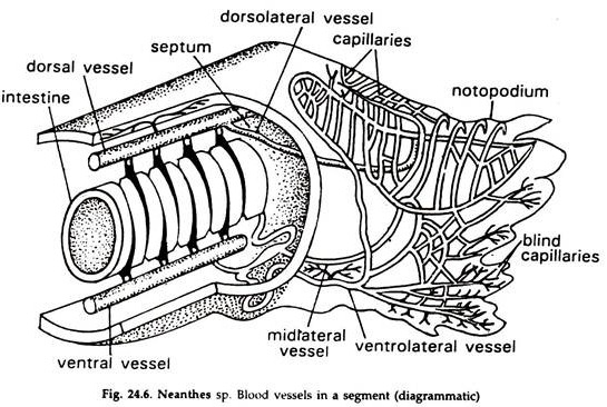
2. There are two main longitudinal vessels (Fig. 24.6) one median dorsal-anal other median ventral to the gut.
Dorsal blood vessel:
A collecting and contractile vessel, carries blood from behind forwards. Anteriorly, it bifurcates and forms a plexus on the oesophagus and joins with the ventral vessel in fifth segment.
i. In stomach region it receives blood from the stomach-intestine by means of two pairs of short dorsointestinals or efferent- intestinals in each segment. It also collects blood from Para podia, nephridia and body wall through lateral vessels. In oesophageal region it supplies blood to oesophagus through branches.
Ventral blood vessel:
A distributing and non-contractile vessel, carries blood from anterior to posterior end. It supplies blood to Para podia, nephridia and body wall and also to stomach-intestine through a pair of short efferent-intestinals in each segment.
The dorsal and ventral vessels are connected on either side in each segment, except the first five, by a pair of loop-like transverse vessels or lateral vessels. Blood from ventral vessel is sent to the skin, Para podia and other organs through afferent vessels from where efferent vessels arise and send blood to the dorsal vessel.
In each segment a pair of ventrointestinal vessels or circumintestinal vessels carry blood from the ventral vessel to the dorsal vessel. The circumintestinal vessel forms a network of capillaries in the stomach-intestine.
A thin sub-neural vessel runs below the -nerve cord, collects blood from the lower body wall and supplies it to the ventral vessel. The bright red colour of the blood is due to the presence of respiratory pigment erythrocruorin, similar to haemoglobin, in solution in plasma. The blood cells remain suspended in the plasma.
Respiratory organs:
Special respiratory organ absent. Gaseous exchange takes place in the surface of Para podia and body wall. Blood is the carrier of oxygen and carbon dioxide.
7. Excretory System of Neanthes:
Annelids tire segmented animals. Their excretion is performed by certain organs known as nephridia. The nephridia with their ducts are usually repeated in each segment. Due to such arrangements, they are also called segmental organs.
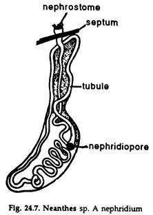
The nephridia of different annelids—though differ considerably in their structural details—are similar in their fundamental construction and extraction of nitrogenous wastes. The nephridia of Neanthes ate simplest (Fig. 24.7). It is a long, narrow, ciliated, folded or coiled tubule, with an opening, the nephrostome, lying in the coelom and a nephridipore at the other end, opening outside.
1. Nephridia are one type and one pair occurs in all the segments except a few anterior and posterior.
2. The body of the nephridium is of an irregular oval shape, the anterior end of which is attached to the mesentery.
3. The body consists of a much coiled ciliated tubule richly supplied with blood.
4. The nephridium passes through the septum and the nephrostome, which is a ciliated funnel, opens in the coelom of the next anterior segment and is a continuation of the tubule of the body.
5. The tubule of the main body opens to the exterior by a fine circular pore, the nephridiopore, capable of dilatation and contraction and is placed on the ventral surface near the base of the ventral cirrus of the Para podium.
6. Wastes, collected by the nephridia are discharged to the exterior directly by the action of the cilia lining the excretory tube.
8. Nervous System of Neanthes:
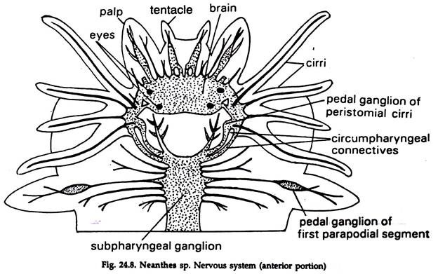
Nervous system is well-developed. It has three components — the central, peripheral and visceral nervous system. (Fig. 24.8).
Central Nervous System:
Cerebral ganglia or brain, circumoesophageal connectives, and ventral nerve cord constitute central nervous system.
a. The brain is a large bilobed mass, situated in the prostomium, above the buccal cavity. Numerous large size neurons or nerve cells and nerve fibres are present.
b. A pair of stout circumpharyngeal connectives arise from the brain, encircle the pharynx and join with the sub esophageal ganglion in the third segment, ventral to the oesophagus.
c. The ventral nerve cord is made up of two solid nerve cords, enclosed in a common sheath. Starting from sub pharyngeal ganglion, in the fourth segment it runs below the ventral blood vessel. The nerve chord is provided with a swelling, formed by a pair of ganglia in each segment.
Peripheral Nervous System:
The nerves emanating directly from the brain and ventral nerve cord to supply different organs are included in peripheral nervous system.
a. The brain gives out four short optic nerves to the eyes, two tentacular nerves, and two palpal nerves to tentacles and palps, respectively.
b. Three pairs of nerves arise from each ganglion of the ventral nerve cord and innervate Para podia, body wall and other structures of that segment.
Visceral Nervous System:
A set of fine nerves arising from the brain and innervating the anterior part of the gut and associated structures constitute visceral nervous system.
Receptors and Sense organs:
Four eyes, two nuchal organs, two tentacles, two palpi, cirri, all located in the prostonium, constitute the sense organs.
Eyes:
The eyes, appearing as black spots, are present on the dorsolateral sides of the prostomium. Each eye is a darkly pigmented cup with a circular aperture, the pupil, a gelatinous lens behind it and a retina occupying the concave surface. The cuticle of the body surface and the epidermis, with somewhat flattened cells, pass over the eye and form the cornea.
The cells of the retina are long and narrow, parallely arranged with one another in a radial direction. The inner-part of the cells are hyaline and project towards the lens; the main body densely pigmented and the outer ends narrow into nerve fibres, forming part of the optic nerve.
Olfactory organs:
The nuchal organs are olfactory in function. They are located near the posterior part of the prostomium on the dorsal side in close contact with the posterior part of the brain. Each nuchal organ has two pits, lined with a ciliated epithelium and gland cells.
Tactile Sense organs:
The tentacles, palpi and cirri are tactile sense organs and help the animals to detect changes in the surroundings.
9. Reproductive System of Neanthes:
1. Neanthes is a dioecious animal. The gonads develop from the ventral coelomic epithelium only in breeding season.
2. In male only one pair of testes lie between nineteenth and twenty-fifth segments.
3. In female, ovaries lie in many segments around blood vessels.
4. The gonads have no ducts; the sex cells are discharged into the coelomic fluid and mature in the fluid.
5. The mature gametes are discharged to the exterior by temporary rupture of the body wall and fertilization take place externally in sea water.
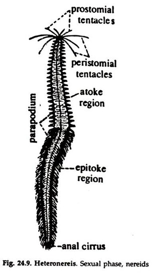
6. Neanthes exhibit polymorphism, a Heteronereis phase develops in the breeding season. Sometimes, during breeding seasons, some individuals leave their usual habitats, the burrows, and become free-swimming. They are called heteronereis.
The body is differentiated into two distinct parts, an anterior atoke and a posterior epitoke (Fig. 24.9):
i. In the atoke eyes increase in size, peristomial cirri become longer.
ii. In the epitoke the Para podia become larger, leaf-like; dorsal and ventral cirri enlarge; additional foliaceous outgrowths are formed; setae become more numerous and longer with oar-shaped blades.
iii. The larger modified Para podia help in swimming and larger body surface serves for rapid respiration during swimming.
iv. The last segment or pygidium develop sensory papillae.
v. The gonads are abundant and confined to the epitoke.
vi. Heteronereis swim upwards and spawn near the surface by rupture of the posterior body wail.
vii. Fertilization takes place in the surface layer of the sea.
10. Development of Neanthes:
The eggs are telolecithal; cleavage spiral; gastrula develops into a trochosphere or trochophore. A larva is a transitional stage in the development of animals. In some animals the eggs, after fertilization, undergo development through a complicated series of stages which are not directly progressive towards the adult stages but assume certain structures which are unlike the parents; although, ultimately, the adult structure is assumed.
The phenomenon is known as metamorphosis and the individual undergoing the process is known as larva. The larva is distinctly separate from the embryo in as much as it is capable of taking its own care and responsibility.
Structure of a Trochophore larva:
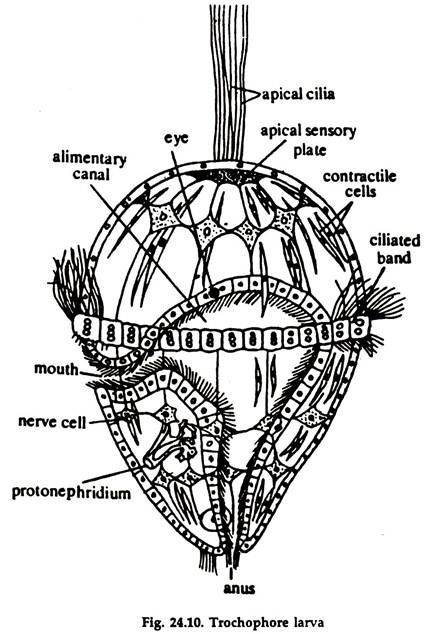
1. The trochophore larva is somewhat conical or oval in shape with a protruding equator, having the general appearance of a spinning top (Fig. 24.10).
2. Externally, it has one-layered epithelium (ectoderm), which is thickened in the apical pole, forming a sensory plate, the apical plate, with a tuft of cilia.
3. Two main bands of cilia encircle the body. The ciliated band around the equator is known as prototroch, which serves as the chief organ of locomotion and food-current guide.
4. The second band, known as metatroch passes below the mouth.
5. In some, a third girdle of a ciliated circlet is present, around the anus. This is known as paratroch.
6. There is no trace of metamerism, the rudiment of the adult trunk is represented only by a small region at the lower pole of the larva.
7. A spacious blastocoel is present, the larva is devoid of coelom at this stage. A pair of protonephridia are present along with mesenchyme and larval muscles.
8. The larva has a complete digestive-tract, ciliated all through, with mouth, stomach and intestine, which opens through an anus at the lower pole.
9. There may be present a ganglionic mass under the apical plate, known as cerebral ganglia.
10. The larval excretory organ consists of a flame cell and an excretory tube, opening to the exterior near the anus.
The trochophore larva is adapted to swimming and feeding on microscopic organisms the plankton. It undergoes successive changes—called metamorphosis—and transformed into adult form.
11. Metamorphosis in Neanthes:
1. With the start of metamorphosis, the posterior end of the trochophore larva beyond prototroch elongates, carrying the gut with it.
2. Traces of segmentation become visible; sometimes a series of segmentation occurs before appearance of Para podia.
3. Gradually, parapodial rudiments with their setae are developed.
4. The external segmentation is accompanied by the division of the mesoderm bands into a series of coelomic sacs.
5. The ventral ectoderm develops a median thickening to form the ventral nerve cords.
6. The larval nephridia, ectodermal muscle bands and ciliated bands of trochophore larva are lost.
7. The region anterior to proto-torch gives rise to prostonium; the cells of the apical plate form the cerebral ganglia; mesodermal tissue grows forward to form a number of coelomic sacs within the prostonium.
8. After settlement, additional segments are added from the growing region.
