In this article we will discuss about:- 1. Description of Scolopendra 2. External Features of Scolopendra 3. Locomotion 4. Glands 5. Digestive System 6. Respiratory System 7. Circulatory System 8. Excretory System 9. Nervous System 10. Reproductive System.
Contents:
- Description of Scolopendra
- External Features of Scolopendra
- Locomotion in Scolopendra
- Glands in Scolopendra
- Digestive System of Scolopendra
- Respiratory System of Scolopendra
- Circulatory System of Scolopendra
- Excretory System of Scolopendra
- Nervous System of Scolopendra
- Reproductive System of Scolopendra
1. Description of Scolopendra:
Scolopendra belongs to class Chilopoda, subphylum mandibulate, phylum Arthropoda. Several species of Scolopendra are found in India. These elongated, swift moving animals live in dark places, burrows, crevices, decaying logs of wood, under partially decomposed leaves in shades of trees. They occur in all tropical and subtropical countries.
2. External Features of Scolopendra:
ADVERTISEMENTS:
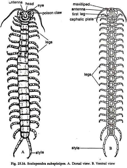
The multiped body is long, worm-like dorsoventrally flattened and divisible into a head and a trunk. The dorsal surface is reddish-yellow and the ventral surface yellowish in colour (Fig 25.16).
Head:
The head consists of head capsule, head lobes and head appendages.
ADVERTISEMENTS:
Head Capsule:
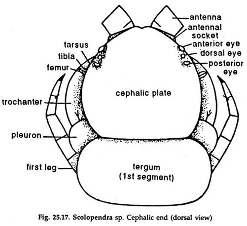
1. It is a somewhat roundish, flattened and well-sclerotized dorsal cephalic plate (Fig 25.17).
2. The cephalic plate bears a pair of antennae in front and four pairs of eyes on the dorsolateral sides.
ADVERTISEMENTS:
3. A pair, each of accessory pleurites and chief pleurites, two pairs of anterior head pleurites, and three pairs of mandibular pleurites form to lateroventral sclerites.
4. Hypo pharyngeal suspensoria constitute the ventral sclerites of the head wall. These support the hypo pharynx.
Head lobes:
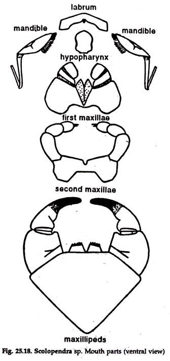
Two median structures, the labrum and the hypo pharynx (Fig 25.18) form the head lobes.
1. Labrum:
It is a preoral median cephalic lobe, the tip being projected posteriorly. The dorsal and ventral sclerites of the labrum are much wider than long and converge to form an extensive blade. The dorsal labral sclerite is provided with numerous sensory setae projecting into the preoral cavity.
2. Hypo pharynx:
It is a tongue-like median lobed structure associated with the ventral wall of the gnathal region of the head and divisible into a long median and two small lateral lobes supported by the hypo pharyngeal suspensoria.
ADVERTISEMENTS:
Head appendages:
The head appendages comprise a pair of antennae, a pair of mandibles, two pairs of maxillae and a pair of maxillipeds (Fig 25.18).
1. Antennae:
Two slender many-jointed appendages narrowing towards the free end.
2. Mandibles:
Two, oriented horizontally on the under-face of the head. Anteriorly, the mandibles are broad and converge laterally while posteriorly they are curved and tapering. The mandible is made up of two units—the posterior unit and the anterior unit. The anterior lobed unit hinges on the posterior unit as a flap. The long incurved sclerite called the posterior sclerite, represents the posterior unit.
The anterior unit is a gnathal lobe whose inner wall is short and membranous and the outer wall is more extensive and sclerotized. The free anterior part of the gnathal lobe bears five large, tricuspid teeth and ventrally crescentic, brush-like setae guarding the oral entrance.
3. First Maxillae:
These are united to form labium-like structure, a dorsoventrally flattened body situated posteroventrally to the hypo pharynx. The coxa is more extensive on the ventral side and bears medially a pair of seta-bearing endities. The telopodite is made of two segments of which the distal is longer and carries sensory setae.
4. Second Maxillae:
These appendage are more leg-like. The coxa is dorsoventrally flattened and is more extensive on the ventral side. The telopodite is movable on its horizontal plane. The tibia bears a dorsal spur while the tarsus carries a comb-like longitudinal row of setae dorsally. Two strong spurs develop on the claw-like pretarsus.
5. Maxillipeds:
Maxillipeds or the poison-claws are virtually the first pair of walking legs modified as accessory feeding organs. Ventrally, the coxae fuse to form a single plate, dorsally they remain as separate plates and anteriorly each bears a pair of dental projects.
The telopodite consists of four segments, trochanter, femur, tibia and tarsus. The poison gland is located in the telopodite and opens at the tip of the claw-like segment which is formed by the fusion of tarsus and pretarsus. Maxillipeds are the first pair of abdominal appendages.
Mouth Parts:
The head lobes and the post antennal head appendages, surrounding the mouth region, form the mouth parts. These consist of mandibles, maxillae, maxillipeds labrum and hypo pharynx. The head being prognathous, the mouth parts are directed anteriorly and under-lap each other from behind forward.
Trunk and Trunk Appendages:
The trunk is segmented and several times longer than the head. It is divided into 24 identical segments.
1. The first trunk segment is apposed to the head and the pair of appendages it bears, are maxillipeds or poison claws.
2. The legs in rest of the segments are seven-jointed, ending in a claw, bearing two ventral spines.
3. The legs become gradually longer posteriorly and the anal legs in the last segment are longest.
4. The telson forming the terminal part of the trunk bears anus and is not considered as a segment.
5. The last segment is the genital segment. In males, a pair of projections—the genital styles, are present in the genital segment.
3. Locomotion in Scolopendra:
The legs support the body in a horizontal position. During locomotion, the legs of one side are lifted and moved forward, the body being supported by the legs of the opposite side. In the next step, the legs are put against the support in a forward position and the legs of the other side are lifted and moved forward. This is repeated and the animal can move very fast.
4. Glands in Scolopendra:
Both unicellular and multicellular glands are present in the hypodermis, below the cuticle.
The glands are of three categories:
1. The poison glands,
2. The salivary glands and
3. The trunk glands.
1. Poison glands:
A poison gland is located in each of the maxilliped.
a. The gland is a somewhat cylindrical sac, lined by numerous unicellular glands.
b. The secretion is carried through a highly cuticularised duct in the middle, opening at the centre of the maxilliped claw.
c. The secretion is yellowish, acidic and helps in defence, lubrication and digestion of food.
2. Salivary glands:
The glands are of three types and help in digestion. All the glands open in the head region:
a. Epipharyngeal glands:
i. Two pairs, located ventral to the brain and open on the epipharyngeal surface through ducts.
ii. The anterior pair is larger and communicate laterally with each other.
b. Labial glands:
i. A pair of lobular glands, located between fifth and seventh segments.
ii. Numerous small ducts arising from the lobules join to form a common duct.
iii. The common duct runs anteriorly along the gut and opens on the ventral surface of the head, between the hypo pharynx and labium.
c. Maxillary glands:
i. One pair, structurally similar to the labial glands and located in the third and fourth segments.
ii. The ducts open on the ventral side of the head region, near the second maxillae.
3. Trunk glands:
Present in the trunk and are of two types:
a. Coxal glands:
One pair in each of the second to fourth segments. The glands are vesicular and their ducts unite to form a common duct opening in the coxopleural plates.
b. Coxopleural glands:
Flask-shaped, many in number and located in the last leg bearing segment. The glands open separately on the coxopleural plates.
5. Digestive System of Scolopendra:
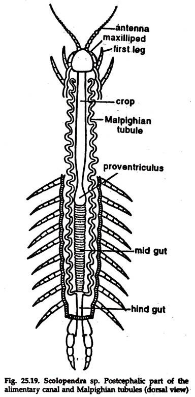
The digestive system consists of a long, straight alimentary canal, starting from the preoral cavity and ending in anus (Fig. 25.19) in the telson.
Salivary glands (previously described) and unicellular glands in the lining epithelium of the gut are digestive glands.
Alimentary Canal:
The alimentary canal is divided into three zones — foregut, midgut and hindgut. The fore and hindgut are lined by cells of ectodermal origin, while the lining of the midgut is endodermal:
a. Foregut:
It is the longest part, extends posteriorly up to 13 leg bearing segment and differentiated into pharynx, oesophagus, crop and proventriculus.
1. Preoral cavity:
The cavity is bounded by the dorsal labral science and membranous epipharyngeal anterodorsally, mandibles laterally and anterior portion of hypo pharynx ventrally. The preoral cavity opens into the pharynx through a buccal chamber.
2. Pharynx:
It is a broad chamber, narrows posteriorly into a short tube, the oesophagus. The internal living epithelium of the pharynx is lined by a cuticular lamina, which is largely folded into a dorsal and-a ventral fold. Ventrally, the spindle-shaped transverse oral muscle marks the anterior-most limit of the pharynx. The extrinsic musculature are seven pairs and act as dilators of the pharynx.
3. Oesophagus:
A short tube ending in crop.
4. Crop:
The longest and thinnest part of the foregut which presents wrinkled appearance due to irregular folds of the cuticular intima internally. Crop extends up to the 12 leg bearing segments.
5. Proventriculus:
A very highly specialised and important part. It is located between the crop and the ventriculus and confined to 12 and 13 segments. Proventriculus is divisible into anterior and posterior proventriculus. The anterior proventriculus acts as a chewing apparatus.
b. Midgut or Mesenteron or Ventriculus:
It extends from the 13 to 19 segment and often bears many superficial transverse constrictions. The loss of cells during secretion (holocrine secretion) is compensated by regeneration of cells out of a group of cells called nidi, embedded in the epithelium.
A protective envelope, the peritrophic membrane, protects the delicate epithelium from injury. The midgut epithelium secreting digestive enzymes are either completely or partially lost (holocrine and apocrine secretion).
c. Hindgut:
It is the posterior ectodermal part of the alimentary canal. The bases of the Malpighian tubules mark the anterior limit of the hind- gut. The extrinsic musculature, extending between anal sclerite and anal passage, helps in evacuation of the excreta through the anus.
6. Respiratory System of Scolopendra:
Respiration is carried by tracheal system (see Cockroach).
Nine pairs of tri-radial, slit-like spiracles are present on the posterior and dorsolateral sides, one in each of the 3, 4, 5, 8, 12, 14, 16, 18 and 20 leg-bearing segments. The spiracles lead to trachaea, which branch profusely and anastomose, ending in tissues
7. Circulatory System of Scolopendra:
In Scolopendra the haemolymph is carried through an open circulatory system formed by blood vessels and sinuses.
Heart and Pericardium Dorsal vessel:
1. The heart, also called dorsal vessel is an elongated, muscular tube, extending from 2 to 21 pedal segments, where it narrows abruptly. In front, it continues as an artery to the cephalic organ.
2. The heart lying in the 3 to 21 segment is enclosed in a cardiac diaphragm or pericardium.
3. The pericardium is drawn into two points in each segment and attached to the tergum of the segment.
4. A pair of openings, the Ostia, communicate the heart with the pericardial cavity in each segment.
5. In each Segment, the heart sends out a pair of vessels to the structures there.
6. Through a number of perforations, the pericardial cavity is in communication with the dorsal sinus.
Ventral vessel:
7. The ventral vessel, also called supraneural vessel, is situated above the nerve cord and extends from 2 to 21 segment.
8. A ring-like stomodaeal vessel connects the dorsal and ventral vessel at the anterior end, which sends four cephalic vessels anteriorly—one dorsal, two laterals and one ventral, supplying haemolymph to the anterior part of the body. Haemolymph is a light- yellow fluid with a few suspended corpuscles, the haemocytes.
Sinuses:
Two large sinuses, dorsal and perivisceral sinus and numerous small sinuses bring back haemolymph from different parts of the body to the heart:
1. Dorsal sinus:
Located dorsally between the pericardium and the body wall. It is in communication with the pericardial cavity through narrow passages and also with the ventral part of the body.
2. Perivisceral sinus:
It is a space around the gut surrounded by a perforated membrane.
Circulation:
Haemolymph from the heart is circulated to different parts of the body by pumping action of the heart. The fluid is finally collected in the dorsal sinus, from where it goes to the heart through the pericardial cavity.
8. Excretory System of Scolopendra:
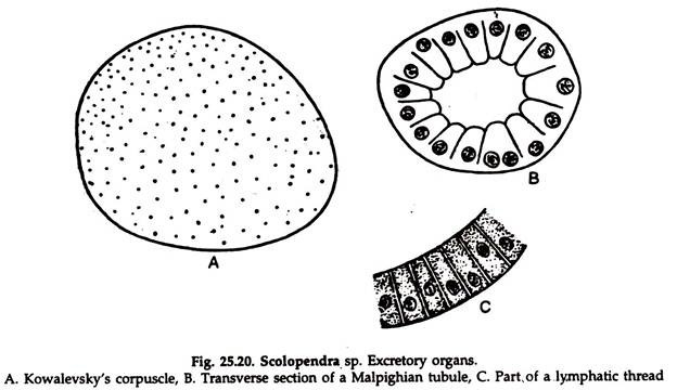
A number of organs (Fig. 25.20) which are concerned with the removal of intracellular wastes from the body and maintenance of a constant internal environment by retaining the necessary constituents constitute the excretory system. The Malpighian tubules are chief excretory organs. Other organs taking part in the process are kowalevsky’s corpuscles lymphatic threads and fat bodies.
Malpighian Tubules:
a. The most important excretory organs are the Malpighian tubules, which are two in number and originate at the junction of the mid and hindgut in the 19th segment.
b. The tubules run posteriorly for a short distance at its beginning and then turn abruptly forward along either side of the alimentary canal, pursuing a sinuous course and reaching as far as the second segment where they terminate blindly (Fig. 25.19).
c. The tubule consists of a columnar epithelium which lacks the striated border towards the lumen. Muscular elements of any sort are absent (Fig. 25.20B).
d. Haemolymph of centipedes contain a considerable concentration of uric acid and the Malpighian tubules eliminate this nitrogenous waste. The uric acid does not crystallize in the tubules. The crystals are formed in the hindgut due to reabsorption of water in the latter. The contents of the tubules are flushed out into hindgut by the continuous circulation of fluid in the lumen of the tubes.
Kowalevsky’s Corpuscles:
The branches of the segmental arteries behind the 3rd segment terminate into somewhat rounded bodies known as Kowalevsky’s corpuscles (Fig. 25.20 A). The corpuscle is a syncytial mass and is responsible for building up blood cells in addition to its excretory function.
Lymphatic Threads:
These are filament-like, bluish in appearance, found round about the Malpighian tubules. Each filament is composed of a row of cells, capable of absorbing carmine particles (Fig. 25.20 C). Fat bodies are supposed to have an excretory function in addition to its storage of reserve materials.
9. Nervous System of Scolopendra:
The central nervous system, peripheral nervous system and visceral nervous system constitute the nervous system of Scolopendra.
Central Nervous System:
It consists of cerebral ganglia, sub-oesophageal ganglia; circumoesophageal connectives, and the ventral nerve cord:
1. Cerebral ganglia or brain:
A pair of ganglia fused to form a bilohed structure, located dorsally in the head.
2. Sub-oesophageal ganglia:
Located in the head, ventral to the oesophagus.
3. Circumoesophageal connectives:
A pair, run ventrally from the cerebral ganglia around the oesophagus to join the sub-oesophageal ganglia.
4. Ventral nerve cord:
It rims posteriorly along the mid-ventral line, up to the posterior end and bears a ganglion in each segment. The genital ganglia lie in the last segment.
Peripheral Nervous System:
The nerves arising from the cerebral ganglia, sub-oesophageal ganglia and ventral nerve cord constitute peripheral nervous system.
1. The cerebral ganglia send nerves to antennae, eyes and other organs in the head.
2. The jaws and maxillipeds are innervated by nerves emanating from sub-oesophageal ganglia.
3. Nerves from the ganglia in the ventral nerve cord innervate organs of the segment concerned.
Visceral Nervous System:
A visceral nerve from the cerebral ganglia innervate the alimentary canal.
Sense Organs in Scolopendra:
Three types of sense organs are present:
1. Eyes (Sensila optica):
One pair of simple ocellus are present on the dorsolateral sides of the head.
2. Sensory hairs and spines:
These are tactile sense organs and scattered all over the body.
3. Organs of Tomosuary:
Located in the base of the antennae and presumably auditory in function.
10. Reproductive System of Scolopendra:
The male and the female can be distinguished from their external dimorphic characters. Males carry a pair of un-segmented genital appendages which are absent in females. Post-genital segment is absent in the females.
Male Reproductive System:
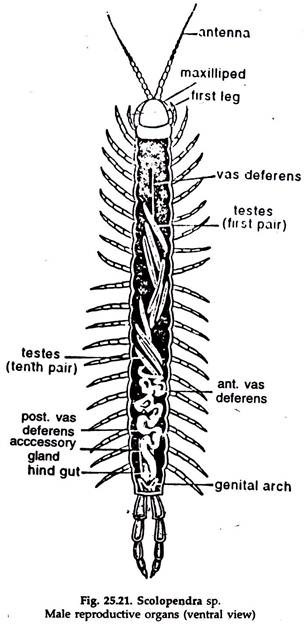
The male reproductive system (Fig. 25.21) consists of testes, vasa efferentia, vas deferens, accessory glands, copulatory organ and the external genitalia. The external genitalia consists of genital, post-genital and anal segments.
1. Testes:
Ten closely juxtaposed pairs of spindle-shaped testes are arranged serially, the anterior end of the first pair being located in the 5th segment. The testis is covered by circular muscle fibres and fibrous tunica of connective tissue. Testis produces two types of sperm cells which differ in size of the head and tail.
2. Vas efferens:
The testis is drawn into a narrow vas efferens at each and. The two vasa efferentia of a pair of testes are bound together in a common fibrous coat and open in a midian vas deferens. The posterior vasa efferentia of a testicular pair join the vas deferens slightly ahead of the anterior ones of the succeeding pair.
3. Vas deferens:
It is differentiated into testicular and post-testicular zones. The testicular part is a narrow tube, concealed under the testes, and it continues as a very thin tube up to the 4th segment where it terminates blindly. The post-testicular part is a fairly transparent and prominent coiled structure.
Anteriorly, it is narrow and highly coiled. Posteriorly, it is dilated at many places for accommodation of spermatophores. More posteriorly it divides into right and left genital ducts which occupy a position ventral to gut. The two then unite to form terminal genital duct communicating with the ejaculatory canal.
4. Accessory glands:
These are two pairs, very closely set, white and lobular exocrine glands. The outer pair is longer and extend forward up to the 18th segment. The ducts of this pair enter the copulatory organ to unite into a common duct, which communicates with the ejaculatory canal while the ducts of shorter pair enter the genital atrium.
5. Copulatory organ:
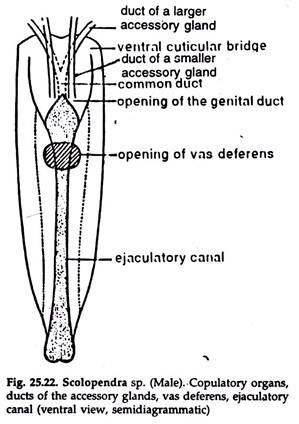
It consists of two vertically placed triangular sclerites united anteroventrally and posterodorsally by cuticular transverse bridge and arthrodial membrane, respectively. It is not a tubular structure but opens ventrally giving the gonopore a slit-like appearance (Fig. 25.22).
Female Reproductive System:
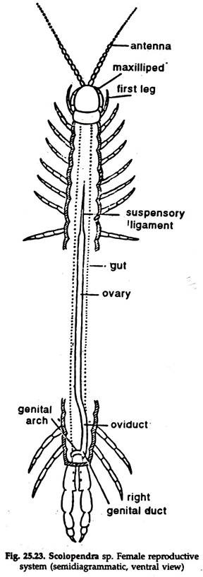
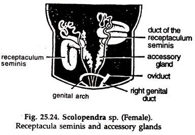
The female reproductive system (Fig. 25.23) consists of an ovary, an oviduct, a pair of receptacula seminis, accessory glands and the external genitalia, the last three being restricted to only genital and anal segments.
1. Ovary:
It consists of an unpaired thin-walled egg tube situated on the mid-dorsal line of the gut beneath the heart. It abruptly narrows down into a terminal filament in the 8th segment and extend up to the 5th segment. The ovarian epithelium is covered by longitudinal striated muscle fibres and connective tissue.
Eggs are produced in the ovary. In the egg, vitelline membrane is apparently lacking although a periplasmic layer is recognisable. The egg contains protoplasmic reticulum, the meshes of which contain yolk globules.
2. Oviduct:
Posteriorly the ovary passes imperceptibly into the oviduct. Intima’ is absent in the inner lining of the oviduct and the epithelium is thrown into folds which permits expansion of the lumen. The muscularis is comparatively thick and composed of longitudinal as well as a few oblique muscles.
The oviduct divides in the last segment into right and left genital ducts. The ducts open separately into the genital atrium ahead of the openings of the ducts of the receptacula seminis and those of the accessory glands.
3. Receptacula seminis:
Two in number, act as sperm storage organs in the last segment of the body (Fig. 25.24). Each receptaculum seminis is a tubular structure, closely bent on itself and communicating independently with the genital atrium by means of a duct which is highly coiled, particularly near the receptacle. The receptacle epithelium produces a secretion which is both protective and nutritive in function.
4. Accessory glands:
A single pair of ectodermal origin, white in colour and lobular in appearance (Fig. 25.24). The ducts of the glands open posteriorly into the openings of the receptacular ducts which are ultimately connected with the genital atrium. They probably help in gluing the eggs by their secretion.
Reproduction in Scolopendra:
The sperms are packed in spermatophores, containing long, thread-like sperms twisted in bundles, and a nutritive fluid. The ovum contains scattered yolk granules. Fertilization and early development internal.
