In this article we will discuss about:- 1. External Features of Ascaris 2. Body Wall of Ascaris 3. Digestive System 4. Excretory System 5. Nervous System 6. Reproductive System 7. Development 8. Larval Wanderings 9. Parasitic Adaptation.
Contents:
- External Features of Ascaris
- Body Wall of Ascaris
- Digestive System of Ascaris
- Excretory System of Ascaris
- Nervous System of Ascaris
- Reproductive System of Ascaris
- Development of Ascaris
- Larval Wanderings of Ascaris
- Parasitic Adaptation of Ascaris
1. External Features of Ascaris:
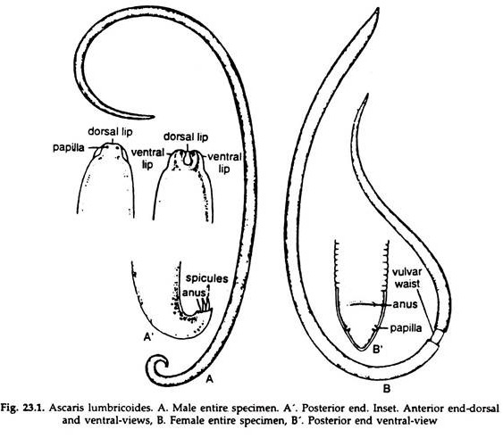
a. The animal is narrow, elongated and light-yellowish brown in colour. Four white longitudinal streaks, one dorsal, one ventral and two laterals are present (Fig. 23.1).
b. The mouth is anterior and terminal, markedly constricted off, and is bounded by three lips; one median and dorsal and two ventrolateral.
c. Excretory pore is ventral and near the anterior end.
d. In male, the tail end is curved ventrally in the form of a hook with a conical tip. The anus opens at this tip and serves as a common aperture for rectum and genital duct. Two copulatory setae protrude from the aperture.
e. In female, the posterior extremity is conical and straight. The anus is situated slightly anterior to the posterior extremity on the ventral surface, guarded by one pair of postanal papillae.
ADVERTISEMENTS:
f. At about one-third of the entire length form the anterior end, the body is narrower in female and this region is known as vulvar waist. The vulva is situated on the ventral surface of the vulvar waist.
2. Body Wall of Ascaris:
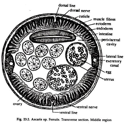
i. The body is covered with a thick elastic transparent cuticle (Fig. 23.2) divisible into several layers with numerous vertical channels. The transverse wrinkles of the cuticle give the characteristic segmental appearance of the body. Cuticle also lines the buccal cavity, oesophagus, rectum, cloaca, vagina and excretory pore.
The cuticle is constituted by the following layers:
ADVERTISEMENTS:
a. Epicuticle:
It is tri-laminate, consisting of two electron-dense layers separated by a less electron-dense layer. Chemically, it is keratin, and also contains quinones and polyphenols.
b. Exocuticle:
Immediately beneath the epicuticle is this layer.
c. Meso-cuticle:
This is a thick layer containing numerous obliquely oriented collagenous fibres.
d. Endocuticle:
This is the innermost fibrillar layer.
ADVERTISEMENTS:
ii. The cuticle remains separated from the underlying tissues by a basal lamina.
iii. The hypodermis or epidermis is a thin syncytial layer under the basal lamina. This layer projects along the mid-dorsal, mid-ventral, and lateral lines and form chords. Excretory canals and lateral nerves run along the lateral chords, while dorsal and ventral chords contain dorsal and ventral nerve, respectively (Fig. 23.2).
iv. In four quadrants, a single layer of muscle cells remain attached to hypodermis. Spindle-shaped muscle cells contain two parts, a muscular and a protoplasmic part. The protoplasmic part remains inwards and connected with the neuromuscular process.
Pseudocoel:
The body cavity is bounded externally by the muscle cells (mesoderm) and internally by the lining of the gut. Embryo logically, it does not develop like a true coelom and is not lined with a layer of mesodermal cells on both the sides. The pseudocoel, in living nematodes, remains filled with a fluid containing mobile phagocytic cells.
3. Digestive System in Ascaris:
The alimentary canal with associated glands constitute digestive system.
Alimentary Canal:
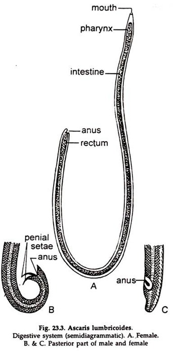
It is a narrow straight tube divisible into three regions (Fig. 23.3) — the club-shaped fore-gut or oesophagus about 10-15 mm long and lined with cuticle; a nanow midgut of endodermal origin and the hindgut or rectum lined by cuticle.
1. Mouth is anterior and terminal and bounded by three lips, one median and dorsal and two ventrolaterals. The mouth leads to a buccal, cavity opening in the pharynx.
2. Oesophagus also called pharynx is a muscular bulb and acts as a suction tube. It leads to intestine.
3. Intestine is a straight tube of one- layered epithelium bounded externally by cuticle.
4. Rectum or the terminal portion of intestine opens to the exterior at the anus.
5. Three branched osophageal glands, one dorsal and two ventrolateral, remain embedded between the muscle fibres.
Feeding and Digestion:
1. Ascaris feeds on hosts intestinal contents, predigested food of hosts, tissues and exudates or blood by the sucking action of the muscular pharynx.
2. Digestion is partly extracellular and partly intracellular.
3. The simplified foods diffuse through the intestinal wall into the fluid filled body cavity and subsequently absorbed by the cells.
4. The excess food may remain stored as glycogen and fat in the epidermal, intestinal, epithelial and muscle cells.
4. Excretory System in Ascaris:
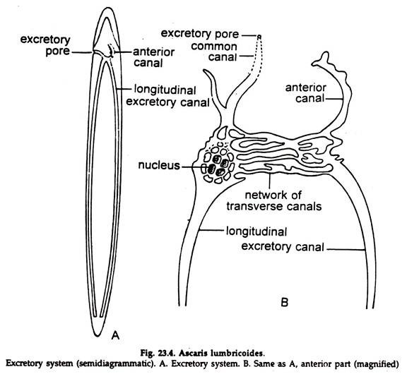
It consist of two lateral longitudinal canals (Fig. 23.4) which meet together ventrally and open in the excretory pore on the ventral surface at the anterior end. Four or six big tuft-shaped cells with ramifying processes are in close contact with the canal. They absorb solid wastes and transfer the same to the canals in a dissolved state.
5. Nervous System of Ascaris:
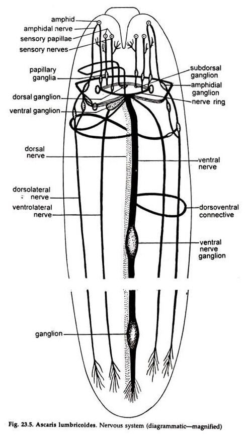
A nerve ring is present around the pharynx from which run six nerves forwards and six backwards (Fig. 23.5). Of the six backwards, two — one dorsal and another ventral — run up to the posterior end and they are connected by transverse nerve bands. The ventral nerve forms a ganglion just in front of the anus.
6. Reproductive System in Ascaris:
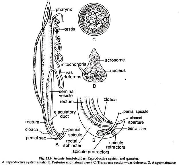
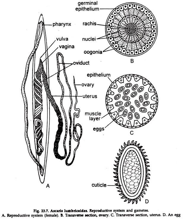
The sexes are separate (Fig. 23.6). Ascaris exhibits considerable sexual dimorphism.
1. In the male, the genitalia (Fig. 23.6) consists of a single, coiled tubule, eight times the length of the body, differentiating into anterior testis, middle region as vas deferens and the posterior region as vesicula seminalis, continuing as ejaculatory duct opening in the anus which is marked by the presence of a pair of penial setae.
2. In the female (Fig. 23.7) ovaries are paired tubes, which pass gradually into oviducts, seminal receptacles and uteri, which join to form an unpaired conical vagina opening in the female gonopore on the ventral surface of the vulvar waist. The female genital organs also remain coiled in the body and, if extended; is found to be several times the length of the body.
7. Development in Ascaris:
The eggs are produced roughly at the rate of 20,000 a day. Fertilization takes place in the upper part of the uterus. Completed eggs being enclosed in chitinoid egg shell pass out of the body of the host with faeces. Development and infection are direct.
Infective embryos reach the intestine of man directly with water, soil, green vegetables, etc. The embryonated eggs move down to the duodenum, where the digestive juice weakens the egg shell. The larva emerges through a rent in the egg shell.
8. Larval Wanderings in Ascaris:
The newly hatched larvae measure about 0.2-0.3 mm and burrow their way through the mucous membrane of the small intestine to reach the lymphatic’s or venules, and from there pass to the liver through portal circulation, where they live for about 3-4 days. From there, they reach the right atrium of the heart and go to the lungs through the pulmonary artery.
Here they leave the blood stream, attack the lung alveoli, and in mass infection the patient develops pneumonic symptoms. In the lungs they moult twice. The larvae now crawl up the glottis and finally reach the intestine again.
The cycle covers, on the average, a period of 10-15 days and the size of the larva increases from 0.2 mm to 2 mm. Fourth moulting occurs in the upper intestine in between 25-29 days and the young continues to grow to attain the normal size. Sexual maturity is attained in about 6-10 weeks-time.
9. Parasitic Adaptation in Ascaris:
Because of certain peculiarities in structure such as the reduction of some organs and specialialisation of others. Ascaris has been adapted to lead a normal life.
1. The body is long, cylindrical in shape with both the ends pointed.
2. The body wall is covered with cuticle, formed of albuminous proteins resistant to the digestive enzymes of the host. Physiological studies show that the worm produces enzyme inhibitors that protect it from the host’s digestive enzyme.
3. Locomotory organs are absent as protection from enemies and food supply are ensured by host.
4. Cilia are completely absent.
5. The alimentary canal is simple and poorly developed as the parasite feeds on the contents of the intestine of host which are in a semi-digested condition.
6. Respiration is almost entirely anaerobic. There is a large accumulation of fatty acids within the parasite, and this settles the first step needed for anaerobic glycolysis of glycogen, richly present in the tissue of the parasite. The energy thus produced is adequate for the worm to carry on its vital processes. It possesses some cytochrome, thus it can respire aerobically also. For this it is sometimes correctly referred to as ‘facultative anaerobe’ and not an obligate anaerobe.
7. Sense organs are poorly developed, being present only on lips in the form of papillae.
8. It is normally hypo-osmotic to intestinal fluid.
9. The reproductive system is highly developed. Numerous eggs are produced and are covered with thick warty chitinous shell which protects them from the digestive enzyme of the host, prolonged dryness and cold for several days.
