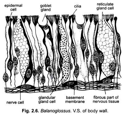
The body wall of Balanoglossus is made up of an outer epidermis, an inner musculature and peritoneum.
Epidermis:
It consists of a single layer of epithelial cells. The epithelial cells are of tall columnar type and have their nuclei near their broader bases.
These cells are mainly of two types:
(i) Ciliated epidermal cells are more numerous and each bears cilia at its free end,
ADVERTISEMENTS:
(ii) Gland cells are lying interspersed between the ciliated epidermal cells.
They are further of three kinds:
(a) Goblet cells are flask-shaped and secrete mucus;
(b) Reticulate cells are long cells with vacuolated cytoplasm which also secrete mucus;
ADVERTISEMENTS:
(c) Mulberry cells are long cells containing coarse cytoplasmic granules and, hence, are also called granular gland cells.
They secrete amylase. The mucus, secreted by gland cells, covers the animal and lines its burrow. The mucus has an obnoxious smell. In addition to these cells, the body wall of proboscis and anterior part of the collar also contain neurosensory cells which take darker stain than the rest. There is no dermis.
Immediately below the epidermis is a thick nervous layer consisting of bipolar and quadripolar nerve cells and fibres which form a network lying in close contact with the epidermal cells. This layer is traversed by the filamentous bases of the epidermal cells that are connected with the basement membrane. The fibres of sensory epidermal cells synapse with the fibres of nerve cells.
ADVERTISEMENTS:
Below the nervous layer is a thick basement membrane made up of two lamellae pressed together. The basement membrane supports the epidermis and serves for attachment of underlying muscles.
Musculature:
The musculature of typical body wall and gut wall is greatly reduced and more or less replaced by muscles arising from the coelomic epithelium. The muscle fibres are smooth and of circular, longitudinal and diagonal types. The muscle layer lies below the basement membrane.
The proboscis musculature comprises a thin layer of circular muscle fibres and a thick layer of longitudinal muscle fibres. The longitudinal muscles fibres obliterate the proboscis coelom and some of the fibres cross one another diagonally. The collar musculature is confined to the collarette and consists of an inconspicuous layer of circular fibres and prominent bands of longitudinal and diagonal fibres. The longitudinal and diagonal fibres, along with connective tissue, also traverse the collar coelom in a criss-cross pattern.
The trunk musculature consists mainly of moderately developed longitudinal muscle fibres which are better developed on the central side. The muscle layer is interrupted by the dorsal and ventral mesenteries and the lateral septa. Several radial muscle fibres are also found in the trunk region. The radial muscle fibres extend between the digestive tract and the body wall and traverse the trunk coelom.
Peritoneum:
It is found just beneath the musculature and outside the coelom. It is also called the coelomic epithelium.
Functions of Body Wall:
(i) The body wall protects the tender internal organs from mechanical injuries,
(ii) Mucus secreted by gland cells adhere sand particles of burrow formed by the animal.
(iii) Foul smell of mucus is protective
ADVERTISEMENTS:
(iv) Neurosensory cells receive and transmit the stimuli.
(v) Musculature helps in locomotion.
