In this article we will discuss about the external morphology of balanoglossus with the help of suitable diagrams.
Shape, Size and Colouration:
The body of Balanoglossus is soft, elongated, cylindrical, bilaterally symmetrical, being richly ciliated all over and covered with mucus. The length of animal varies from 2 cm to 2.5 meters according to species. Most forms are drab coloured, though reddish tints are present, several species are luminescent due to mucus. They have an offensive odour.
Division of Body:
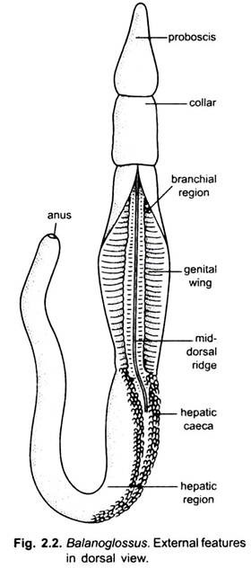
The body is un-segmented and divided into three regions, viz., proboscis or protosome, collar or mesosome, and trunk or metasome (2.2).
Proboscis:
ADVERTISEMENTS:
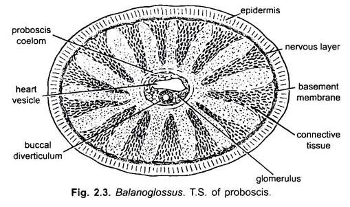
The proboscis forms the anterior part of the body and is either rounded or conical in shape. It is continued posteriorly into a short, narrow neck or proboscis stalk which is attached with the collar. The proboscis is hollow and has thick muscular walls. Its cavity the proboscis coelom opens to the outside by means of a small opening called the proboscis- pore situated mid-dorsally near its base.
In certain cases there are two proboscis-pores. In some species the proboscis-pore does not communicate with the outside environment, but terminate blindly. Beneath the proboscis stalk, the base of proboscis lies in a U-shaped ciliated epidermal depression, the pre-oral ciliary organ.
The proboscis sits in the collar somewhat like an acorn in its cup, a character that has given the name “acorn worms” to the group. The mouth, which is always wide open and incapable of closing completely, lies on the ventral side and its lips are the ventral edges of the collar region.
Collar:
The collar lies posterior to the proboscis and anterior to the trunk. It is a short cylinder usually about as wide as long and mostly shorter than the proboscis although sometimes longer. The funnel-like anterior part of the collar, the collarette, embraces the proboscis stalk and usually also the posterior part of the proboscis. Posteriorly the collar is sharply demarcated from the trunk by a circular indentation.
ADVERTISEMENTS:
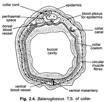
The surface of the collar is often marked with elevations, depressions, and specially circular grooves. The collar is also muscular and possesses two coelomic cavities, called the collar coelom. The right and left coelomic cavities are separated from one another by dorsal and ventral mesenteries. The coelomic cavities of collar are completely cut off from the proboscis cavity.
The collar cavity as well as the proboscis cavity is crossed by numerous strands of connective tissue which give the region a spongy appearance. The collar cavity communicated with the exterior by a pair of short ciliated collar tubes (canals) leading into the first pair of gill-pouches.
Locomotion:
The functional significance of the cavities and water pores in the proboscis and collar may best be explained through a description of the burrowing habits. When on the surface of the sandy bottom Balanoglossus pushes the tip of the proboscis into the sand, moving it around by muscular contractions until a shallow, cylindrical hole is made. Then the proboscis empties its water content through its pore and collapses. This allows the collar to enter the hole.
By taking in water through the pores the collar expands so as to fit tightly into the hole like a cork in a bottle. The well-filled collar then gives a point of resistance for further rooting movements of the refilled proboscis, which loosens sand and stores it into the scoop-shovel mouth.
Then both proboscis and collar relax and the latter squirms deeper into the hole before tightening its hold again. Once the collar gets a firm grip, the animal makes rapid progress and soon buries itself the tail end is left near the surface, and at intervals comes out and deposits a pile of castings somewhat after the fashion of earthworms.
Trunk:
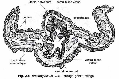
The trunk is the elongated posterior part of the body. It is somewhat fiat and annulated on the surface. It has a mid-dorsal and a mid-ventral longitudinal ridge along its entire length for the accommodation of corresponding nerve and blood vessel. The trunk is divisible into three parts- an anterior branchiogenital region, a middle hepatic region, and a posterior abdominal or post-hepatic region.
(i) Branchiogenital Region:
On the dorsal surface of the branchiogenital region of the trunk is a double row of small pore—the branchial apertures, one on either side of mid-dorsal ridge. Each row is situated in a long furrow. These pores increase in number during growth. In some species the most anterior are overlapped by a posterior prolongation of the collar called the operculum.
The branchiogenital region has a pair of lateral, thin, flat and longitudinal flaps. In these the genital ridges, gonads are situated. In some genera, the genital ridges are so prominent that they form a pair of wing-like lateral folds, the genital wings, but in other genera folds are absent.
(ii) Hepatic Region:
ADVERTISEMENTS:
The hepatic region is dorsally marked externally with irregular elevations due to sacculations produced by projecting hepatic caeca of the intestine along with body wall.
(iii) Abdominal Region:
The abdominal region is longest and cylindrical. It tapers gradually and has a terminal anus. The coelom of the trunk is divided into two lateral closed cavities by vertical partition between the body wall and gut-wall.


