Digestive system consists of an alimentary canal and digestive glands.
Alimentary Canal:
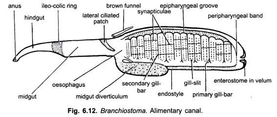
The alimentary canal is a straight tube from mouth to anus. It is lined by ciliated epithelium. It is divisible into mouth, buccal cavity, pharynx, oesophagus and intestine opening by an anus.
1. Mouth:
Mouth lies anteroventrally beneath the rostrum. It is a large oval aperture bordered by the membranous oral hood. The oral hood is formed of anterior lateral projection of trunk.
ADVERTISEMENTS:
2. Buccal Cavity or Vestibule:
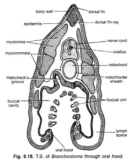
The mouth leads into a space enclosed by the oral hood lined by ectoderm called the buccal cavity or vestibule which is funnel-shaped. The free margin of the oral hood is beset with ten to eleven pairs of oral cirri or oral tentacles. Their number increases with the age and varies from animal to animal. These cirri are ciliated and also bear sensory papillae. The margin of oral hood and cirri are supported by stiff gelatinous skeletal rods.
The oral cirri remain converged inward to form a sieve over the mouth at the time of feeding. Thus, they check the entry of larger particles of food into the vestibule. At the back of vestibule is a vertical membranous velum having a hole in the middle known as enterostome.
The enterostome is also called a mouth. Since the enterostome leads into an endoderm-lined pharynx and not into an ordinary ectoderm-lined vestibule or stomodaeum, it cannot correspond to the mouth of chordates. Hence, the front opening into the vestibule is a true mouth.
ADVERTISEMENTS:
(i) Muller Organ:
The epithelium (ectoderm) of the oral hood is projected to form six to eight pairs of finger-like folds, each having a ciliated groove borded by ciliated ridge. These folds are collectively known as wheel organ or Muller’s organ or rotatory organ. Outer of these, the mid-dorsal groove is the largest and ends in a pit or depression in the roof of buccal cavity. This is called Hatscheks groove and Hatscheks pit respectively. Both are ciliated and glandular, and secretes mucus.
(ii) Velum and Enterostome:
ADVERTISEMENTS:
At the posterior end of the buccal cavity is present a circular, membranous velum. It is centrally perforated by the enterostome which leads into pharynx. The velum has circular muscles which act as sphincter for opening and closing the enterostome. The edges of the velum bear 12 or more slender, ciliated velar tentacles which normally project backwards forming a strainer.
3. Pharynx:
The pharynx is a large, laterally compressed sac occupying nearly one-half anterior part of the body. It is suspended in the atrial cavity which encloses it from all sides except the dorsal.
(i) Pharyngeal Wall and Gill-Slits:
The lateral walls of the pharynx are perforated by more than 150 pairs of gill-clefts which bear no gills. The gill-clefts are metameric, vertical apertures when first formed in the larva, but each gets divided into two. New gill-clefts are added with age at the posterior end of the pharynx; hence, their number varies in different specimens. Between the gill-clefts the wall of the pharynx is known as gill-bars or gill-lamellae.
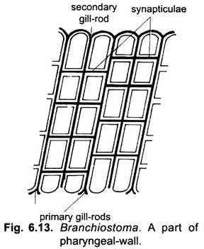
The pharyngeal gill-bars are of two types- primary gill-bars and secondary gill-bars or tongue-bars. They alternate regularly and differ in their structure and mode of development. A primary gill-bar is formed of the tissue between two successive gill-clefts after they have perforated to the exterior.
It is composed of the wall of the pharynx and the body wall of the larva. The secondary gill-bars arise as down growths of the dorsal wall of larval gill-clefts, the wall grows downwards dividing the original gill-cleft into two halves vertically.
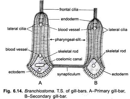
Both primary and secondary gill-bars are covered on their external or outer surface by sparsely ciliated ectodermal or atrial epithelium, but on their inner, anterior and posterior surfaces by endodermal pharyngeal epithelium which is heavily ciliated.
The cilia on the anterior and posterior endodermal surfaces of gill-bars are long and called the lateral cilia which propel water, and the cilia on the inner endodermal surface of each gill- bar form a long but narrow tract of frontal cilia which propel mucus. In the middle each gill- bar has a mesodermic core of connective tissue, blood vessels and gill-rods.
The gill-bars are supported internally by gelatinous skeletal gill-rods. All gill-rods are united dorsally, but ventrally their free ends are forked in the primary gill-bars, and unforked or simple in the secondary gill-bars. The primary gill-bars are connected to each other by transverse junctions called synapticula which also contain gelatinous rods and blood vessels. The synapticula develop only after the gill-clefts are completed, they make the pharynx look like a basket, somewhat like the branchial sac of urochordates.
The primary gill-bars contain a narrow coelomic canal running throughout the length of each and communicating dorsally and ventrally with other corresponding coelomic spaces, they also have three blood vessels in each, running lengthwise. The secondary gill-bars with simple gill-rods have no coelom and only two blood vessels run through each of them.
(ii) Epipharyngeal Groove:
Besides the ciliated gill-bars, there are other ciliated tracts in the pharynx. Running mid-dorsally is a ciliated epipharyngeal groove which leads into the opening of oesophagus at the posterior end of the pharynx.
(iii) Endostyle:
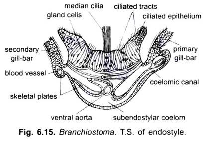
In the mid-ventral wall of the pharynx is a shallow groove called endostyle. The endostyle is lined with four longitudinal tracts of glandular epithelial cells which secrete mucus. These tracts are separated by ciliated epithelial cells of which the median row of cells bears very long cilia. The endostyle is supported below by the two gelatinous skeletal plates.
Beneath these is present the subendostylar coelomic canal enclosing the ventral aorta. A similar endostyle is found in urochordates and in the ammocoete larva of lampreys, but the larval endostyle is lost during metamorphosis of lamprey, it contributes to the formation of a thyroid gland in the adult. Like thyroid gland of vertebrates, it also concentrates radioactive iodine.
(iv) Peripharyngeal Bands:
The epipharyngeal groove and endostyle are joined to each other anteriorly by two narrow ciliated peripharyngeal bands. Each running on the lateral side wall of the pharynx behind the velum and demarcates a narrow anterior prebranchial region of the pharynx from the proper large posterior pharynx. It is devoid of cilia and gill-slits.
4. Oesophagus:
The pharynx opens posteriorly into a straight narrow ciliated oesophagus followed by a wide midgut or intestine.
5. Intestine:
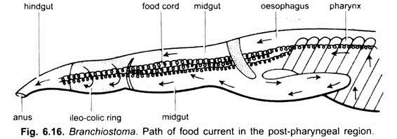
Intestine is a long tube suspended in the atrium by the dorsal mesentery arising from the dorsal body wall. It is continued into a narrow hindgut opening by an anus lying slightly to the left where the caudal tin begins. Arising ventrally from the junction of oesophagus and midgut is a blind pouch called midgut diverticulum which extends in front on the right of the pharynx. The entire gut is suspended from the dorsal body wall by a dorsal mesentery. The gut has a lining of epithelial cells and a thin covering of smooth muscles.
The gut has several internal ciliated tracts in its lining, there is a crescent-shaped lateral tract of cilia in the midgut which directs food into the midgut diverticulum; there is a dorsal tract in the midgut; between the midgut and the hindgut is a large ciliated ileo-colonic or ileocolic ring which chums food. In the hindgut is another dorsal tract of cilia.
Digestive Glands:
The midgut diverticulum is often called liver or hepatic caecum, but it does not resemble a liver in structure or function, though its blood vessels are somewhat like those found in the liver of vertebrates. The midgut diverticulum is a digestive gland and is comparable to a vertebrate pancreas.
Physiology of Digestion:
Food:
Branchiostoma feeds on protozoans, diatoms, algae, desmids and other organic particles suspended in sea water.
Feeding:
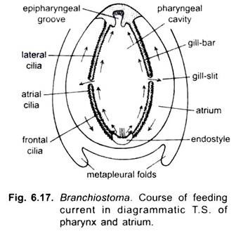
Branchiostoma is a ciliary feeder. Action of cilia of the pharynx causes a current of water containing food. The current of water enters the mouth and goes to the pharynx from where it passes through gill-clefts into an atrial cavity, and then it goes out through an aperture of the atrial cavity called atriopore. In feeding the oral hood is extended and oral cirri are turned inwards, they prevent sand from entering the mouth.
Rotary movements of cilia of wheel organ direct water towards the pharynx, but some food particles fall out of the water current, they are mixed with mucus secreted by groove of Hatschek and passed back into the pharynx. When the main water current passes through the enterostome into the pharynx, mainly due to lateral cilia of gill-bars, the suspended food particles fall on the gill-bars where they get entangled in mucus secreted mainly by the endostyle and to some extent by the pharyngeal epithehum.
Mucus secreted by endostyle is transferred to the lateral wall of the pharynx by its lateral rows of cilia; the median row of long endostylar cilia keeps supplying mucus to the lateral rows of cilia of the endostyle. These cilia lash outwards throwing the muccus on the lateral wall of pharynx.
The frontal cilia of gill- bars beat along the length of the bars in such a way that they propel mucus from the lateral to the mid- dorsal side of the pharynx. In this way a stream of mucus with food particles passes from the lower- side into epipharyngeal groove. The cilia of epipharyngeal groove beat backwards moving the cord of mucus and food into the oesophagus.
The peripharyngeal bands also collect and pass to the epipharyngeal groove any food particles which fall out of the water current at the extreme anterior end of the pharynx (where no gill-clefts are present). Food and mucus are not transferred from the endostyle into the peri-pharyngeal bands, as is often stated.
The cord of food and mucus passes down the gut by action of cilia. It is moved from the oesophagus into the midgut where a lateral tract of cilia directs it into the midgut diverticulum, from here the cord is returned again into the midgut. The iliocolic or iliocolonic ring rotates the cord of food causing the food and enzymes to mix, and then the cord of food is moved into the hindgut.
Digestion:
Digestive enzymes are secreted by the epithelial cells of the gut and midgut diverticulum. They are mixed with food as it passes along. Digestion starts in the midgut and is continued in the hind-gut.
Besides this extracellular digestion, intracellular digestion also occurs in which food particles are taken into the epithelial cells of the hindgut and digested there. Some papillae on the floor of the atrium contain phagocytic cells which engulf food particles which may pass into the atrial cavity. Absorption of digested food takes place mostly in the hindgut and to lesser extent in the midgut.
