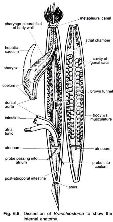
In this article we will discuss about the external morphology of branchiostoma with the help of suitable diagrams.
Shape, Size and Colour:
Branchiostoma is about 3.5 to 6.0 cm in length. The body is slender, somewhat translucent, superficially fish-like, laterally compressed and pointed at both ends, hence, the name lancelet. The anterior two-thirds of the body is roughly triangular in transverse section. The posterior third is nearly oval in section. The pointed anterior end of trunk projects in front as a snout or rostrum. Below the rostrum is an oral hood formed by dorsal and lateral projections of the body. It bears twenty or more stiff oral cirri or tentacles.
Body Divisions:
The body is distinguished in two regions- the trunk and tail. The true head is lacking.
Fins:
Branchiostoma has three unpaired fins- dorsal, caudal and ventral. Paired fins are absent. Along the mid-dorsal side of the body is a dorsal fin running along the whole length. It is joined to a somewhat broader caudal fin around the tail. Mid-ventrally is a ventral fin running from the caudal fin to an atriopore.
The dorsal fin is supported by a series of fin rays, and nerves the ventral fin by a series of paired fin rays. The fin rays are made of a gelatinous substance and connective tissue. Fin rays are lacking in caudal fin. It is important to note that the fins do not have the same structure as the fins of fishes.
Folds:
The oral hood is continuous posteriorly with two lateral membranous hollow metapleural folds running ventrally up to the atriopore. Both the folds are joined by a horizontal fold of body wall, the epipleur. It forms the floor of the atrium internally.
Myotomes and Gonads:
ADVERTISEMENTS:
The body wall shows metameric segmentation, being made of <-shaped blocks of muscles called myotomes or myomeres. These are found just beneath the transparent body wall. Beneath the myotomes on both sides are present a series of gonads between mouth and atriopore.
Apertures:
Below the pointed anterior extremity is a large median aperture, the mouth surrounded by a frill-like membrane, the oral hood. Its edge is beset with numerous tentacles or cirri, immediately in front of the anterior end of the ventral fin, and partly enclosed by the metapleural folds, is a rounded aperture, the atriopore. A short distance from the posterior extremity of the body (trunk) is the anus, placed asymmetrically on the left side at the base of the caudal fin. The post-anal portion of the body is called the tail.
Musculature:
The muscles present beneath the body wall exhibit metameric segmentation. Muscle layer consists of a large number about 60 – 62 of muscle segments or myomeres or myotomes, separated from one another by partitions of dense connective tissue, the myocommas.
They have the appearance, in a surface view, of a series of very open “V”s with their apices directed forwards. The myotomes have striated muscle fibres lying longitudinally; they are inserted at each end into myocommata. Contractions of these segmental myotomes cause side to side bending of the body by which the animal swims.
The myotomes of two sides of the animal do not correspond but are placed alternately. On the dorsal side the myotomes are very thick enclosing the nerve cord and notochord, this is characteristic of vertebrates also, but in many invertebrates the muscles are of uniform thickness all around. Ventrally between the metapleural folds in the floor of atrial cavity are present a special set of transverse muscles in the anterior two-thirds of the body. No such muscles are seen in vertebrates.


