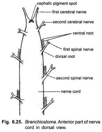
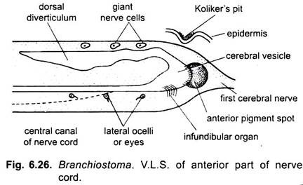
In Branchiostoma, nervous system is simple, having no brain as found in higher chordates.
Nerve Cord:
There is a mid-dorsal, hollow, tubular neural tube or nerve cord lying above the notochord. It starts behind the anterior end of the notochord and tapers to a point posteriorly and ends a short way in front of the hinder end of the notochord. The neural tube contains a narrow central canal or neurocoele throughout its length and filled with cerebrospinal fluid.
Nerve cells are grouped around the central canal, and fibres arising from nerve cells are placed superficially, while in invertebrates the nerve cells are superficial and nerve fibres are placed centrally. Like annelids, the neural tube on the dorsal side has some giant neurons with giant nerves fibres running lengthwise.
Anteriorly the central canal dilates within the cerebral vesicle forming its ventricle. It gives out a dorsal diverticulum which extends behind over the central canal for some distance. The cerebral vesicle has two receptor organs, a pigment eye spot in its anterior wall and an infundibular organ on its floor.
Nerves:
From the anterior part of the neural tube (cerebral vesicle) arise two pairs of sensory nerves going to the oral hood cirri and sense organs. These nerves are called cerebral nerves and lack ventral roots. The body behind the cerebral vesicle gives off spinal nerves in segmental pairs.
Each spinal nerve has a dorsal root with afferent sensory fibres entering, and a ventral root made of several separate efferent motor fibres leaving the neural tube. The dorsal roots come from the skin and is of mixed type (i.e., sensory and motor) whereas, the ventral roots are motor and go to myotomes.
The dorsal and ventral roots do not unite to form a mixed spinal nerve, as in vertebrates. Moreover, a dorsal root of a spinal nerve is slightly posterior to its ventral root. The dorsal roots also contain fibres running to the wall of the gut. The spinal nerves of the two sides do not correspond; the dorsal roots of one side are opposite the ventral roots of the other side. These spinal nerves have no myelin sheath, they are primitive.
Autonomic Nervous System:
There is an autonomic nervous system having two nerve plexuses in the smooth muscles of the wall of the intestine. These are connected to the neural tube by visceral nerves (preganglionic) through the dorsal roots. The autonomic nervous system is apparently represented only by the parasympathetic system; it controls the unstriped muscles of the gut.
Sense Organs (Receptors):
Amphioxus is richly supplied with simple receptors.
1. Eye Spots or Ocelli:
Simple eyes or ocelli are rows of black pigmented spots along the entire length of the ventro-lateral sides of the neural tube. They are arranged in definite tracts and are sensitive to light. Each eye has an outer pigment cell or melanocyte and a lens-like photosensitive cell with a striated apical border or vitreous body which secretes a pigmented cap. It acts as a lens. The photosensitive cell receives one nerve fibre from the nerve cord. The light-sensitive eyes are responsible for orientation of the animal as it burrows in sand.
ADVERTISEMENTS:
2. Pigment Spot:
Pigment spot is a large pigmented spot in the extreme anterior wall of the cerebral vesicle. It lacks the structure of an eye and has no sensory function, But when the animal burrows with its anterior end projecting, then the pigment spot shields the eyes from frontal stimulation by light and probably acts as a thermoreceptor.
3. Infundibular Organ:
Infundibular organ is a depression in the floor of the cerebral vesicle. It is lined by long ciliated epithelial cells. Internally it gives out a Reissner’s fibre which enters into the neurocoel. It detects changes in the pressure of the fluid in the neural tube.
4. Kolliker’s Pit:
Kolliker’s pit or olfactory pit is a pocket of ciliated ectoderm cells above the cerebral vesicle slightly to the left side. It marks the position of neuropore by which the neural tube opens anteriorly in the larva, but the neuropore closes in the adult. Kolliker’s pit is probably not an olfactory organ since it has no specialised sensory cells, nor it has any connection with the brain.
5. Papillae:
Papillae are groups of sensory cells on oral cirri and velar tentacles. They are sensory to touch. The cells of the velar tentacles and oral cirri are also chemoreceptors having gustatory and olfactory functions.
6. Sensory Cells:
ADVERTISEMENTS:
Sensory cells are found scattered all over in the epidermis, they are specially abundant on the dorsal side of the body and on oral cirri. Each sensory cells has a nerve fibre at its lower end and at the outer end it has a hair-like sensory process projecting from the cuticle. These cells respond to contact and some of them are concerned with determining the nature of the sand in which the animal will burrow, for the animal avoids sand which is too fine.
The above mentioned receptors in or near the integument are collectively known as exteroceptors or external receptors.
7. Free Nerve Endings:
Besides the above external receptors, there are sensory nerve endings in myotomes which respond to internal stimuli, such as muscular contractions. These internal receptors in muscles are called proprioceptors.

