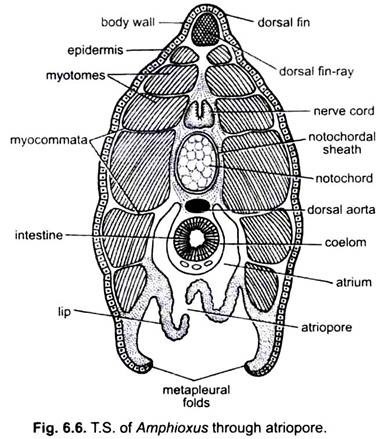
In Branchiostoma, exoskeleton is not found. But a few endoskeletal structures are present which are not cartilaginous or bony.
Notochord:
There is a cylindrical and tapering notochord at both the ends, remarkable for its extension from the tip of the rostrum to the end of the tail. It lies immediately above the alimentary canal and between the left and right myotomes. It is formed of large alternate fibrous and gelatinous, vacuolated cells and having the nuclei confined to its dorsal and ventral regions. Around these cells is a structure less layer, secreted by the cells, enclosed in a notochordal sheath of connective tissue.
Notochordal sheath is produced dorsally into an investment for the canal enclosing the central nervous system. The turgid cells and notochordal sheath give the notochord its stiff elasticity enabling it to prevent any shortening of the body when myotomes contract. In Branchiostoma alone among chordates, the notochord extends anteriorly in front of cerebral vesicle. This prolongation is probably an adaptation to its habit of burrowing rapidly into the sand.
Oral Ring:
The oral hood is supported by a gelatinous ring of cartilaginous consistency made of separate rod-like pieces lying end to end, and corresponding in number with the cirri. Each piece gives out a rod forming the axis of an oral cirrus.
Gill-Rods:
Gill-bars of the pharynx and endostyle are supported by skeletal rods made of elastic, gelatinous substance. Gill-rods are composed of agglutinated elastic fibres. These will be discussed along with the pharynx.
ADVERTISEMENTS:
Fin-Rays:
Dorsal fin is supported by a single row of fin-rays, while the ventral fin has a double row of supporting fin-rays. These fin-rays are made of connective tissue containing a gelatinous substance inside. Fin-rays are separated from one another by small cavities.
