The blood vascular system includes the heart, blood vessels (arteries and veins) and blood.
Heart and Pericardium:
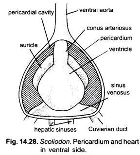
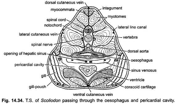
The heart of Dogfish (Scoliodon) lies mid-ventrally beneath the pharynx in the head region. It is a simple dorso-ventrally bent S-shaped muscular tube. It lies in the pericardial cavity, bounded by a two-layered membranous pericartium. It is a median triangular cavity lying between the gills with the apex directed forwards, and is almost completely occupied by the heart. The heart of Dogfish (Scoliodon) contains only the impure blood, hence, it is called venous or branchial heart.
The heart consists of four chambers:
(a) The sinus venosus,
ADVERTISEMENTS:
(b) The atrium,
(c) The ventricle and
(d) The conus arteriosus.
(a) Sinus Venosus:
ADVERTISEMENTS:
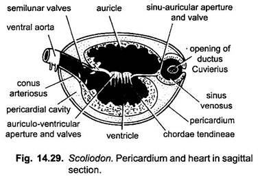
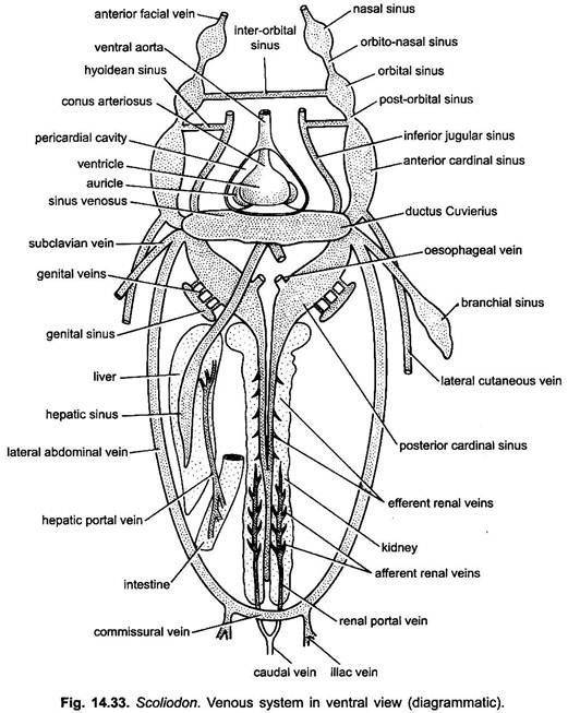
The sinus venosus is a triangular, thin- walled posterior chamber extended transversely and lying fused along the base of the pericardial cavity. Laterally it receives two large veins, the ducti Cuvieri, one on each side, while two hepatic sinuses open into it in the postero-median line. The sinus venosus opens anteriorly into the atrium through the sinu-atrial sinu-auricular aperture, guarded by a pair of membranous valves, the sinu trial valves. These valves prevent the backward flow of the blood.
(b) Auricle:
ADVERTISEMENTS:
The atrium (auricle) is a large triangular sac, lying in front of the sinus venosus and dorsally to the ventricle. It occupies the dorsal half of the pericardial cavity. Its walls are somewhat thicker than those of the sinus venosus. Its lateral posterior angles produced into processes which project laterally at the sides of the ventricle like ears it opens into the ventricle through the atrio-ventricular aperture, guarded by a bilabiate valve which prevents the backward flow of the blood.
(c) Ventricle:
The ventricle is the most prominent pear-shaped chamber of the heart It has very thick muscular walls because it propels the blood to the entire body. The inner surface of the ventricle is produced into numerous muscular strands which give it a spongy texture.
The opposite walls are held in place by chordae tendineae and also protect the ventricle to expand beyond its capacity. It communicates dorsally with the atrium through the atrio-ventricular aperture and anteriorly with the conus arteriosus.
(d) Conus Arteriosus:
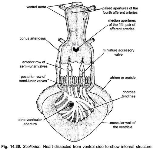
The conus arteriosus is a stout muscular tube which arises from the ventricle and extends up to the anterior end of the pericardial cavity. Its inner wall is provided with two transverse rows of semi-lunar valves, each row containing three valves, one dorsal and two ventro-laterals in position. In addition to these, there is always a small accessory valve on either side of the dorsal valve. The anterior valves are larger than the posterior valves.
The free-ends of the valves are connected to the walls of the ventricle by fine tendinous threads to keep the valves in position. These valves prevent the backward flow of blood into the ventricle. The conus arteriosus is continued forward through the wall of the pericardium as the ventral aorta. The ventricle and conus constitute the forwarding pump for the blood.
ADVERTISEMENTS:
Working:
The heart of dogfish is venous type since it receives only venous or deoxygenated blood. The blood also flows through the heart only once (single circulation). Heart pumps the venous blood into the gills for aeration. For this purpose, the different chambers of the heart rhythmically contract at regular intervals in a definite sequence—first sinus venosus → auricle → ventricle → conus arteriosus. Contraction of the heart is called systole and relaxation is called diastole. The valves at different places prevent the backward flow of blood into the preceding chambers. The walls of the heart are also supplied oxygenated blood through coronary vessels.
Arterial System:
Arteries have more muscular walls than the veins. The heart pumps the blood into the arteries which are of following types:
1. Afferent Branchial Arteries:
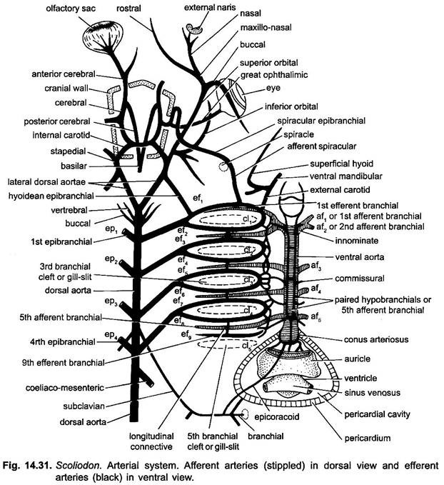
The ventral aorta arises from the conus arteriosus and runs forwards along the ventral surface of pharynx right up to the posterior border of the hyoid arch, where it bifurcates into two short branches, the innominate arteries. Each innominate artery divides into first and second afferent branchial arteries. The first afferent branchial artery runs along the posterior border of hyoid arch and supplies branches to all the gill-lamellae of the hyoidean demibranch.
The second afferent branchial artery runs along the posterior border of the first branchial arch and supplies arterial branches to the anterior and posterior gill-lamellae of the first branchial arch. The third, fourth and fifth afferent branchial arteries arise independently from the ventral aorta, almost equidistant from one another but the fifth branchials of both sides arise from a common median aperture from ventral aorta.
These run along the outer borders of the second, third and fourth branchial arches. They supply the blood to the third, fourth and fifth gill-arches respectively. These all break up into capillaries in the gill-lamellae.
2. Efferent Branchial Arteries:
The blood from the capillaries of gill-lamellae is collected by a series of blood vessels called the efferent branchial arteries. There are nine efferent branchial vessels on each side-pretrematic is along the anterior border of gill-pouch and the post-trematic along the posterior border of gill-pouch. Of these the first eight join in pairs to form four complete loops around the first four gill-pouches.
The ninth runs along the anterior border of the fifth gill-pouch. The four loops and ninth efferent branchial artery are joined by short longitudinal connectives running across the interbranchial septa. The loops are further connected with one another by a network of longitudinal commissural vessels between their ventral extremities. From the upper end of each of the four efferent branchial loop arises an epibranchial artery. These run backwards and inwards to the mid-dorsal line, and all the four pairs of epibranchial arteries unite in the middle in the roof of the pharynx to form the median dorsal aorta. The half-loop of ninth efferent branchial has no epibranchial of its own.
3. Arteries of the Head:
The first efferent branchial or hyoidean efferent and a small part from the dorsal aorta supply blood to the head. The first efferent branchial vessel or hyoidean gives rise to three branches-the external carotid, afferent spiracular and hyoidean epibranchial.
(а) The external carotid artery arises from the antero-ventral comer of the first efferent branchial vessel and runs along the outer surface of the hyoid arch. It divides into two branches– (i) The ventral mandibular supplying the coraco-mandibular muscles and muscles of lower jaw, and (ii) Superficial hyoid supplying the skin and sub-cutaneous tissues over the ventral part of the hyoid arch.
(b) The afferent spiracular artery arises at about the middle of the hyoidean efferent. It runs forward on the outer side of the hyomandibular and surrounds the spiracle by the spiracular epibranchial artery across the floor of the orbit and then enters the cranium through a small foramen. Just before entering the cranium it gives off the great ophthalmic artery to the eye-ball. Within the cranium it unites immediately with a branch of internal carotid to form the cerebral artery which is a short vessel dividing immediately into anterior and posterior cerebral arteries.
(c) The hyoidean epibranchial artery arises from the dorsal part of the first efferent and runs forward and inward. At the level of the orbit it receives a branch from the dorsal aorta and then divides into a stapedial and an internal carotid, (i) The stapedial artery runs forward and enters the orbit where it gives two branches, one supplying the eye-muscles and the superficial tissues in the region above the auditory capsules and other supplying the anterior boundary of the orbit. The former is called the inferior orbital, while the latter is called the superior orbital, (ii) The internal carotid passes inward along a groove in the roof of the buccal cavity and enters the cranium through a foramen.
Within the cranial cavity it divides into two branches one of which unites with its fellow of the opposite side, while the other unites with the stapedial to form the anterior and posterior cerebral arteries.
4. Dorsal Aorta and its Branches:
The dorsal aorta is formed by the union of four pairs of epibranchial arteries. It runs backwards along the whole length of the body, lying beneath the vertebral column in the trunk. In the tail region it continues within the haemal canal of the tail vertebrae as the caudal artery. The dorsal aorta supplies oxygenated blood through its paired and unpaired branches to all the structures behind the gills.
It gives off the following branches from anterior to posterior sides:
(i) Lateral Aortae:
These arise from the anterior end of dorsal aorta and divides into two. Each branch unites with the hyoidean epibranchial of its side.
(ii) Buccal:
A pair to the roof of buccal cavity.
(iii) Vertebral:
Paired vessels to the vertebral column.
(iv) Subclavian:
Close to the union of the fourth epibranchial arteries, the dorsal aorta gives off a pair of small vessels, the subclavian arteries. Each subclavian passes outwards and backwards to the pectoral girdle and the pectoral fin of one side, which it supphes.
(v) Coeliaco-Mesenteric:
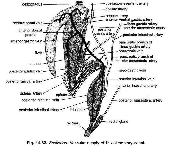
It is a large median artery arising from the dorsal aorta slightly behind the junction of the fourth pair of epibranchial arteries. It divides into two unequal branches- the smaller coeliac and the larger anterior mesenteric. The coeliac artery supplies the stomach and liver, etc., and the anterior mesenteric artery supplies the pancreas, intestine and the rectum.
(vi) Lieno-Gastric Artery:
It arises from the dorsal aorta a short distance behind the origin of coeliaco-mesenteric. It is also a median blood vessel giving off branches supplying the genital organs, stomach, spleen and posterior part of the intestine.
(vii) Posterior Mesenteric Artery:
It is a small median vessel which supplies the posterior end of the gonads and finally ends in the rectal gland.
(viii) Parietal Arteries:
These are a series of paired vessels arising at intervals along the whole length of the dorsal aorta and supplying the body wall. The renal arteries are small paired vessels arising from the parietal arteries and supplying the kidneys.
(ix) Iliac Arteries:
These are a pair of arteries similar to the parietals, each of them extending into the pelvic as a femoral artery and there breaks up into capillaries.
5. Hypobranchial Blood Plexus:
It includes a pair of longitudinal median hypobranchial arteries present in the ventral wall of ventral aorta. These are connected with each other by transverse vessels. Four commissural vessels connect hypobranchial with the lateral hypobranchial chain of its side.
The lateral hypobranchial chain of either side is connected with the ventral ends of the efferent branchial loops. Both the hypobranchial arteries unite posteriorly to form a median coracoid artery which divides into a coronary artery for heart and a pericardial artery for pericardium.
Venous System:
In Dogfish (Scoliodon), the venous blood from the entire body is returned to the heart by the veins. The veins differ in structure from the arteries in possessing thin walls and frequently contain valves which prevent backward flow of blood. Many veins form wide irregular spaces without definite walls called sinuses during their course.
It is characteristic of venous system of Chondrichthyes (elasmobranchs).
The venous system of Dogfish (Scoliodon) can be divided into the following systems viz.:
(i) Anterior cardinal system,
(ii) Posterior cardinal system,
(iii) Hepatic portal system, and
(iv) Cutaneous system.
1. Anterior Cardinal System:
The anterior cardinal system collects blood from the part of the body which lies anterior to the heart, i.e., head region.
It consists of:
(i) A pair of large vessels or sinuses the internal jugular veins. Each internal jugular comprises the olfactory sinus, orbital sinus, post-orbital sinus and the anterior cardinal sinus. The olfactory sinus receives blood from the rostral region by an anterior facial vein.
The orbital sinus lies in the orbital region and is connected with its fellow of the opposite side by an inter-orbital vein. The orbital sinus opens into the post-orbital sinus and finally into the anterior cardinal sinus.
The anterior cardinal is a large sinus and lies in between the dorsal ends of the gill-pouches and the muscles of the body wall. It opens into the ductus Cuvieri behind and receives a hyoidean sinus from inferior jugular sinus. It also receives blood from the five dorsal nutrient branchial sinuses of the gills.
(ii) A pair of inferior jugular sinus. These are small median ventral sinuses, one on either side. These collect blood from the bucco-pharyngeal region, gill-pouches and heart region and then open laterally into the ductus Cuvieri.
2. Posterior Cardinal System or Renal Portal System:
The posterior cardinal system collects blood from the part of the body which has posterior to the heart. It consists of a median caudal vein, two renal veins and two large posterior cardinal sinuses.
(i) The caudal vein runs below the caudal artery and lies in the haemal arch of the caudal vertebrae. It receives numerous branches on either side from the myotomes of the tail. On entering the abdominal cavity dorsally to the cloaca, the caudal artery bifurcates into two branches, the right and left renal portal veins,
(ii) The renal portal veins continue forward along the dorso-lateral margins of the kidneys and give out many afferent renal veins which break up into capillaries in the kidney. The renal portal veins receive small parietal veins from the body-wall. The blood from the sinusoids of the kidneys is collected by the efferent renal veins which join together to form a single vessel.
Anteriorly it divides into two posterior cardinal sinuses. The two posterior cardinal sinuses lie close together along the roof of the abdominal cavity. In the region of the oesophagus, the posterior cardinals expand into wide thin-walled sacs and each receives an oesophageal vein from oesphagus and numerous genital veins from the genital sinus. Finally, each posterior cardinal opens into the cuvierian duct by a relatively small aperture opposite the opening of the anterior cardinal sinus.
3. Hepatic Portal System:
The blood from the alimentary canal and its associated glands is collected by a number of veins which unite to form the hepatic portal vein. The hepatic portal vein is formed by the union of the anterior and posterior intestinal veins.
The large vein so formed receives a lineo-gastric vein, and anterior and posterior gastrics and then runs forward to divide into two branches which enter the right and left lobes of the liver. The blood brought to the liver is distributed in the lobes of the liver by fine capillaries. It is collected by a second set of capillaries which together form two large thin-walled sinuses, the hepatic sinuses which open into the sinus venosus by two small apertures.
4. Lateral Abdominal Veins:
There are two lateral abdominal veins. Each collect blood from body wall, cloacal region and paired fins of its own side. In the posterior region both are connected by a commissural vein and each receives an iliac vein from the pelvic fin formed by the union of an outer femoral and inner cloacal vein. Each lateral abdominal vein anteriorly receives a subclavian vein from pectoral fin and then empties into the ductus Cuvierius laterally.
Lateral vein instead of joining with the subclavian, opens separately into the precaval. The genital sinus of either side opens into the posterior cardinal sinus of their own side.
5. Cutaneous System:
The cutaneous system includes a mid-dorsal, a mid-ventral, and two lateral cutaneous veins. The dorsal cutaneous vein runs beneath the skin along the mid-dorsal line and collects blood from the skin of the dorsal side.
The ventral cutaneous vein runs along the mid-ventral line beneath the skin and joins the lateral abdominal in front of the cloacal vein behind. The lateral cutaneous vein runs along each side just beneath the lateral line canal. Each lateral cutaneous empties its blood into the branchial vein in front.
Blood:
The blood of Scoliodon, consists of a colourless plasma and corpuscles in it Corpuscles are of two types- RBCs (erythrocytes) are oval and nucleated bodies and contain respiratory pigment, haemoglobin and WBCs (leucocytes) and amoeboid cells resembling with lymphocytes of other vertebrates.
