The endoskeleton is composed entirely of cartilage hardened at places by calcifications. The true bones are entirely absent.
The endoskeleton can be divided into:
(i) Axial skeleton comprising the vertebral column, the skull and
(ii) Appendicular skeleton including the pectoral and pelvic girdles and the skeletal elements of fins.
Axial Skeleton:
ADVERTISEMENTS:
Vertebral Column:
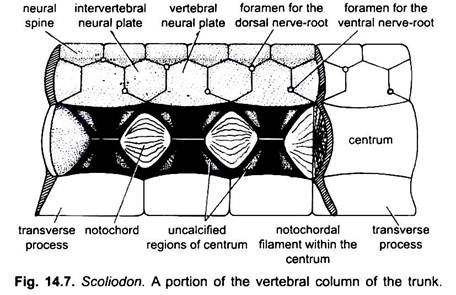
The vertebral column comprises a chain of about 130 vertebrae, which develop around an elastic axis of vacuolated cells, the notochord. The vertebral column is clearly divisible into two regions- trunk and tail.
1. Trunk Vertebra:
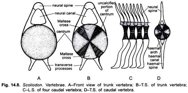
A trunk vertebra consists of ventral, cylindrical acentrum surrounding the slender notochord, a dorsal neural arch enclosing the neural canal for the spinal cord, and a pair of transverse processes projecting ventro-laterally from the centrum.
ADVERTISEMENTS:
The vertebrae of trunk region are amphicoelous, i.e., the centrum bears concavities on anterior as well as posterior surfaces. In transverse section the centrum shows four wedge-shaped calcified fibro-cartilages, arranged in the form of a cross which is called Maltese cross.
These cartilages also leave some uncalcified area between them, hence the vertebra or centrum is known as asterospondylus. Through the series of centra are found remnants of the notochord. The concave anterior and posterior surfaces of the centra are covered by a dense calcified layer. The centrum bears on its dorsal side two neural arches which meet mid-dorsally to form the neural spine.
Each neural arch consists on each side a small cartilage the neural plate (basidorsal) which becomes completely fused with the neural process in the adult between successive neural plates is present a series of interdorsal or interneural plates Small median cartilages, the neural spines fit in between both neural and inter-neural plates of opposite sides and form the complete arch.
ADVERTISEMENTS:
From the ventrolateral side of the centrum there arise a pair of slender transverse processes which are directed outwards. The transverse processes of the anterior vertebrae are short and extend outward, but in the posterior trunk vertebrae they bend downward and enclose an open groove.
2. Caudal Vertebra:
The caudal vertebra is slightly modified in comparison to the trunk vertebra. The neural arch is short, while the neural spine is very much large and pointed. The transverse processes converge to meet and fuse together forming a haemal arch which encloses a haemal canal through which caudal artery and vein pass.
The haemal arch is produced into a long haemal spine pointing backwards. The haemal spines of the posterior caudal vertebrae support the large ventral lobes of the caudal fin, while the dorsal lobe is supported by neural spines. The posterior part of the vertebral column is bent dorsally to form an asymmetrical or heterocercal tail.
The trunk vertebrae are monospondylous but caudal vertebrae are diplospondylous, there being two complete vertebrae to each mytome.
Significance of Vertebral Column:
The vertebral column forms the axial scaffolding of the body and serves as a foothold for the insertion of all the body and tail muscles concerned in the locomotion. The segmentation increases the flexibility of the column but the vertebrae are so closely bound to one another by strong fibrous connective tissue that there is a little freedom of movement for individual vertebrae. In fishes flexion of the column as a whole is all that is necessary for prevailing lateral movements of the body.
Skull:
ADVERTISEMENTS:
The skull of Dogfish (Scoliodon) consists of:
(i) The cranium surrounding and protecting the brain,
(ii) The sense capsules closing the olfactory and auditory organs, and
(iii) The visceral skeleton forming the jaws and the skeletal supports of the pharynx and gills.
Cranium and Sense Capsules:
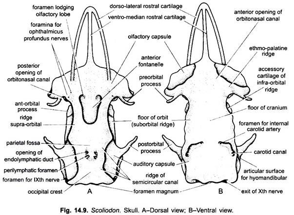
The cranium is simple and cartilaginous. Its general shape is very much like that of a violin box, open in front and behind, with an arched roof and a flattened floor. Two pairs of sense capsules, the olfactory and auditory, are intimately fused to it. In the hinder region the massive auditory capsules project on either side into a deep concavity for lodging the membranous labyrinth.
The anterior region, besides presenting swelling on each side is formed by the olfactory capsule. Anteriorly the cranium gives off three cartilaginous bars which are prolonged forward and unite to form rostrum.
The cranium may be roughly divided into four regions:
(a) The occipital region,
(b) The auditory region,
(c) The orbital region and
(d) The ethmoidal region.
(a) Occipital Region:
The occipital region forms the posterior part of the cranium. It is perforated behind by a large median opening, the foramen magnum through which the brain is continued into the spinal cord. Just below the foramen magnum there is a cup-shaped concavity which encloses the notochord and lies opposite to the concave centrum of the first vertebra.
On either side of the foramen magnum, there is a prominent rounded occipital condyle for articulation with the first vertebra. Above the foramen magnum, the roof of the occipital region carries a prominent median crest called the occipital crest. External to the occipital condyle, on either side lies a large foramen for the exit of the tenth or vagus nerve.
(b) Auditory Region:
The auditory region lies in front of the occipital region and comprises two auditory capsules enclosing internal ears and the part of the cranium with which they are intimately fused. The auditory capsules remain separate from the cranium in the embryo but become firmly united with it in the adult. On the roof of the cranium, in between the two auditory capsules there is a marked oval depression, the parietal fossa.
The parietal fossa possesses two pairs of apertures. The anterior pair of apertures are smaller through which the endolymphatic duct of each internal ear pierces the cranium. The posterior pair is comparatively larger and forms the openings of the perilymphatic spaces of the two capsules. Each auditory capsule possesses three ridges which mark the position of three semicircular canals of the internal ear. Hyomandibula of hyoid arch (2nd) articulates with in the post-orbital groove of both sides.
(c) Orbital Region:
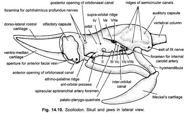
The orbital region lies anterior to the auditory region almost in the middle of the cranium. It is hollow, called the orbit and lodges the eyes, their muscles, the large orbital blood sinuses and a large number of nerves. Each orbit is bordered dorsally by a supra-orbital ridge or crest, ventrally by infra-orbital or sub-orbital ridge, anteriorly and posteriorly by pre- orbital and post-orbital processes respectively.
These processes are the extensions of supra-orbital ridge. Within the orbit in the cranial wall are present a number of foramina for cranial nerves. The anterior boundary of the orbit is formed by a posteriorly directed slender process which arises from the posterior part of the olfactory capsule and is called the pre-orbital cartilage.
A similar process arising from the anterior part of the auditory capsule directed forwards marks the posterior boundary. This process is known as post-orbital cartilage. A lateral outgrowth of the floor of the orbit forms the suborbital ridge which is produced anteriorly into the ant-orbital process forming a place for the insertion of ligaments of the upper jaw.
(d) Ethmoidal Region:
The ethmoidal region consists of two olfactory capsules and the rostrum. The lateral olfactory capsules are large oval cartilaginous cups at the anterior end of the skull which are firmly united to the cranium in the adult. Each capsule lodges an olfactory sac. Postero-ventral surface of each capsule has a short, stout ethmopalatine ridge.
A thin median cartilage, the internasal septum or mesethmoid separates the two olfactory capsules from each Other. The cranial roof in this region shows a depression, the anterior fontanelle covered over by a sheet of connective tissue. Within each olfactory capsule there is a large opening leading into the cranial cavity, through which the olfactory nerve enters the olfactory sac of its own side.
The rostrum or snout is formed by the fusion of three cartilaginous bars. In front of the anterior fontanelle arise a pair of dorso-lateral cartilages, one from the roof of each olfactory capsule. These cartilages run forward to converge and meet in front with a median ventral cartilage which projects from the base of the cranium.
The floor of the cranium is broad and flat and bears towards its hind end two obliquely transverse grooves called the carotid canals. The anterior half of each carotid canal bears two apertures, one leading into the cranium transmitting the internal carotid artery and the other leading into the orbit transmits the stapedial artery. Immediately behind the olfactory capsules lie two large prominent articular surfaces for the attachment of the ethmopalaline ligaments of the upper jaw, while in front of these articular surfaces lie the anterior openings of the orbitonasal canals.
Foramina of the Skull:
A number of foramina for nerves and blood vessels perforate the skull wall. On each side of the anterior fontanelle are apertures for ophthalmic branches of V and VII nerves. Each olfactory capsule has a large foramen for the olfactory nerve. The wall of the orbit on each side has a foramen for the optic nerve, and exits or foramina for the oculomotor, trochlear, trigeminal, abducens and the facial nerves.
In front of the foramen for trigeminal nerve is a small foramen for the entrance of the spiracular epibranchial artery. Behind the auditory region is a large foramen on each side for the glossopharyngeal nerve. On the outer side of each occipital condyle is a large foramen for the vague nerve. The floor of the skull in the posterior part of the cranium has two large carotid canals, each has two foramina, one of the Internal carotid artery, and the other for the stapedial artery.
Visceral Skeleton:
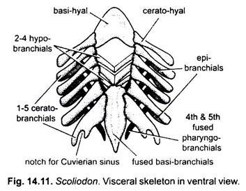
The visceral skeleton or the splanchnocranium is closely connected with the cranium and forms the jaws and pharyngeal skeleton. It consists of a series of seven pairs of U-shaped visceral arches which lie in the sides and floor of the pharynx.
(i) The first visceral arch or mandibular arch, forms the upper and lower jaws bearing teeth. The upper jaw or palatoquadrate has two cartilages joined in front. Near its anterior end it has an orbital process from which ligaments arise to join the cranium. The lower jaw has two Muckel’s cartilages united anteriorly. At its posterior end each Meckel’s cartilage has an articular surface for a movable joint with the upper jaw.
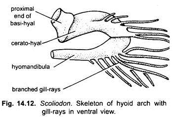
(ii) The second visceral arch or hyoid arch is made of five cartilages, a lower median basihyal in the floor of the pharynx supporting the buccal floor and tongue, and a pair of ceratohyal in the buccal floor and a pair of dorsal hyomandibular which bear slender cartilaginous branched gill-rays. Each hyomandibular is connected at its upper end articulates with the cranium, and at its lower end with the palatoquadrate (upper jaw) and Meckel’s cartilages (lower jaw). The jaws are not directly joined to the cranium but are only suspended by the hyomandibular, this type of suspensorium is hyostylic and is found in most Chondrichthyes. The remaining hyoid arch supports a gill.
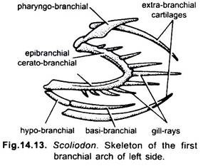
(iii) Branchial arches- The remaining posterior five visceral arches are called branchial arches because they support the gills and the wall of pharynx allowing muscular movements of the pharyngeal wall for a respiratory current. Typically each branchial arch is made of nine pieces of cartilages, a lower median rod-like basibranchial to which are attached on each side a ventra hypobranchial, a lateral ceratobranchial, and epibranchial, and a dorsal pharyngobranchial cartilages. The ceratobranchials and epibranchials bear slender cartilaginous unbranched gill-rays which support the gills. Besides these there are four pairs of extra-branchial cartilages which lie at right angles to the first four pairs of branchial arches.
Variations in the Branchial Arches:
In Dogfish (Scoliodon) some of the cartilages are lost or fused with others. It has all five basibranchials fused to form a single cartilage. The hypobranchials are very small in the first branchial arch and absent in the fifth. All five ceratobranchials are present, the fifth one being very large. The first four epibranchials are present but the fifth one is lost. There are four pharyngobranchials, those of the fourth and fifth branchial arches having fused into a single piece.
The skull is, thus, formed from the chondrocranium, first visceral arch, and the dorsal part of the second visceral arch. This condition is found in all fishes and tetrapoda, except in ungulates and primates in which the middle part of the hyoid arch also fuses with the skull in the auditory region as the styloid process.
The skull forms the support of the head with protective coverings for the brain and sense organs, and it forms the skeleton of the jaws and internal support of the pharynx and the gills. The presence of jaws is a great advance in evolution, they support the mouth which is used not only in feeding and biting but it is also used for handling the environment, as in nest building in many fishes it is useful for holding objects.
Appendicular Skeleton:
The appendicular skeleton includes the median and paired fins and the pectoral and pelvic girdles. Of the median fins there are two dorsals (anterior and posterior) and a ventral and a caudal. The paired fins include the pectoral and pelvic fins.
1. Median Fins:
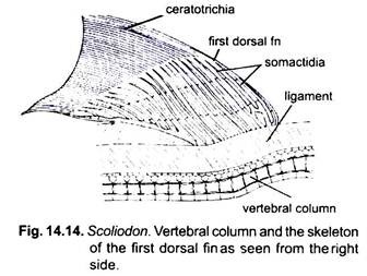
The skeleton of two dorsals and the median ventral fins consists of a series of cartilaginous rods called somactids or pterygiophores bearing distally a double series of numerous horny fin-rays or ceratotrichia. A wide strip of ligamentous tissue connects the somactidia of the fin with the vertebral column. Each somactid is divided typically into three segments, the proximal, mesial and distal segments. The segments of the adjoining somactidia tending to fuse at their bases.
The first dorsal fin is supported by twenty-one somactidia, the proximal segments of which are fused along the posterior border of the fin to form an obliquely horizontal axis in this region. The second dorsal fin and median ventral or anal fin are built on the same plan as the first dorsal. In the caudal fin there are no somactidia but the neural and haemal spines of the vertebral column are elongated and flattened to support the dorsal and ventral lobes of the fin.
Pectoral Girdle and Fins:
(i) Pectoral Girdle:
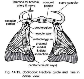
The pectoral girdle (Fig 14.15) is embedded in the lateral and ventral body wall posterior to gills and has two curved rods of cartilage fused together in the middle below the pericardium. Each half is a scapulocoracoid cartilage, its upper curved part is the scapula and the lower part is the coracoid. At the dorsal end of scapula is a distinct suprascapular part. In between the scapula and coracoid is a glenoid region with three foramina for branchial artery and nerve. The girdle is not connected to the vertebral column.
The glenoid region articulates with the pectoral fin by a triple facet. The pectoral girdle provides a fulcrum for attachment of muscles which move the fin, the muscles attached to coracoid control the visceral arches thus they help in breathing and swallowing.
(ii) Pectoral Fin:
Pectoral fin is supported by a series of both endoskeletal and exoskeletal elements. The endoskeleton has pterygiophores cartilages to which muscles are attached for moving the fin. The proximal pterygiophores are basals and distal ones are radials There are three thick basals, an outer propterygium, a middle mesopterygium, and an inner metapterygium Extending from the basals are three rows of numerous radials, i.e., proximal, mesial, and distal radial cartilages collectively called somactidia. Arising from distal somactidia are horny, unjointed exoskeletal ceratotrichia or fin rays. They support the fin blade.
Pelvic Girdle and Fins:
(i) Pelvic Girdle:
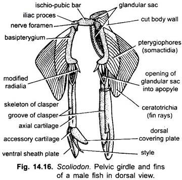
Pelvic girdle (Fig. 14.16) is a flat transverse rod of cartilage called ischiopubic bar. It lies transversely with its ends bent, near each end is a nerve foramen. On each side of the girdle is an acetabular region for attachment of basal cartilage of pelvic fins. The pelvic girdle is more primitive than the pectoral girdle. In fishes it is never connected to vertebral column and lies embedded in the abdominal wall in front of cloaca.
(ii) Pelvic Fin:
Like pectoral fin, the pelvic fin also has the same endoskeletal and exoskeletal structures. There is a single curved basal cartilage called the basipterygium to which several somactidia (radials) are attached. The fin blade is supported by exoskeletal ceratotrichia or fin rays attached to somactidia.
The pelvic fin shows sexual dimorphism, in the male a part of each fin is modified to form a clasper or myxopterygium. It arises from the distal end of the basipterygium. It is an elongated tubular axial cartilage which is dorsally grooved. At its distal end is a sharp pointed style enclosed within a dorsal and a ventral plate and at the upper end of the style is a small accessory cartilage.
The clasper has an anterior aperture, the apopyle which communicates with cloaca and the siphon and a posterior hypopyle opening on a sharp, pointed style. The siphon lies beneath the skin of the ventral surface of the body and communicated with the claspers by means of paired ducts. The claspers are covered with skin. They are erectile and used for transporting sperms during copulation.
The skeleton of median fins is similar to that of paired fins. The endoskeletal pterygiophores form a series of radials (somactidia) which bear a double series of distal exoskeletal ceratotrichia. In the caudal fin there are no somactidia, their place being taken by neural and haemal spines of caudal vertebrae.
