In Scoliodon, the exoskeleton comprises the placoid scales, embedded in the skin.
Structure of a Placoid Scale:
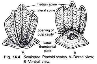
A typical placoid scale consists of mainly two parts- the basal plate and the flat trident spine. The basal plate is diamond-shaped and lies embedded in the dermis and firmly placed by Sharpey’s and other connective tissue fibres. The basal plate is formed of the trabecular calcified tissue closely allied to the cement. The inner surface of the basal plate bears an opening which leads into the pulp cavity.
During life, the pulp cavity is filled with vascular connective tissue called the pulp containing numerous odontoblasts (dentine-forming cells), blood vessels, nerves, and lymph channels. The spine is flat and trident and projecting out of the skin and backwardly directed.
The surface of the spines is not smooth but presents a stratified appearance under the microscope. The spine is composed of a hard calcareous substance, the dentine. The dentine is traversed by minute nearly parallel canaliculi with delicate branches containing long protoplasmic processes of odontoblasts.
ADVERTISEMENTS:
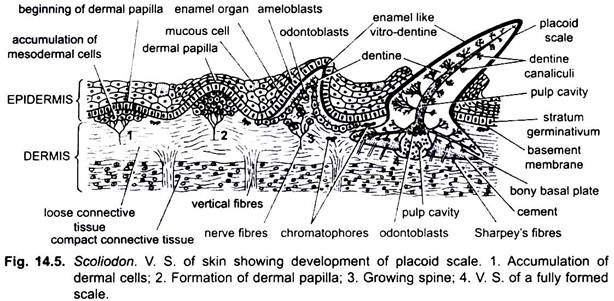
It is coated externally with a layer of hard shiny enamel-like vitrodentine secreted by ameloblasts of the overlying ectoderm. The placoid scales are derived partly from the dermis and partly from the epidermis. The basal plate and the dentine of the spine are derived from mesoderm, while the enamel is secreted by the ectoderm.
Development of Placoid Scale:
A group of mesodermal cells of dermis collect in the dermis just beneath the epidermis to form a dermal papilla. The outermost cells of dermal papilla become the odontoblasts. The dermal papilla grows upwards pushing the epidermis, then it takes the shape of a basal plate and spine. The odontoblasts secrete dentine all around forming the basal plate and spine of the placoid scale. The dentine of the basal plate becomes calcified.
ADVERTISEMENTS:
The Malpighian layer of the epidermis overlying the dermal papilla is now called the ameloblasts form the enamel organ, which form the vitrodentine over the dentine of the spine. The dermal papilla in a growing scale forms pulp in the pulp cavity of the scale. The overlying epidermis moves away and also wears off so that the spine projects above, while the basal plate remains embedded in the dermis.
Homology of Placoid Scale:
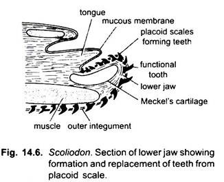
Placoid scales are the forerunners of vertebrate teeth because the two have essentially the same form, structure and embryonic development. The gradation from placoid scales to teeth is seen in the mouth of a shark. Shark teeth are enlarged placoid scales formed in the skin of jaws and are, thus, homologous with placoid scales.
But there are objections to this supposition, and a more recent view is that both placoid scales and teeth are modified remnants of the bony dermal plates found in the ancestral ostracoderms and placoderms, so that teeth and placoid scales are homologous structures. Moreover, it has been shown that there is no epidermal enamel layer in placoid scales, but it is a layer of vitrodentine formed by mesodermal cells.
Enamel is the hardest substance in the body, whereas vitrodentine is only a hardened outer layer of dentine. The enamel organ does not secrete enamel, it only plays a role in shaping the spine, so that the entire placoid scale is mesodermal like the scales of bony fishes, whereas a tooth has an enamel covering derived from ectoderm.
ADVERTISEMENTS:
The claim that a graduation from placoid scales to teeth is observed in the mouth of a shark is interpreted as a divergence shown between vitrodentine-covered placoid scales, on the one hand, and enamel-covered teeth on the other.


