In this article we will discuss about the nervous system of dogfish with the help of suitable diagrams.
Central Nervous System:
The central nervous system is essentially tubular and encloses a narrow central canal within the spinal cord which widens out anteriorly to form the ventricles of the brain.
1. Brain:
The brain of Scoliodon is large and considerably advances to that of the Cyclostomata. It is divided into a forebrain, a midbrain and a hindbrain.
ADVERTISEMENTS:
(i) Forebrain:
The forebrain comprises the cerebrum and the diencephalon.
(a) Cerebrum:
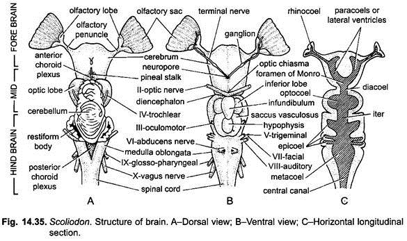
The cerebrum forms the anterior part of the brain and is an undivided mass of nervous tissue. There is no median dorsal groove to separate the cerebrum into right and left cerebral hemispheres. Its roof and floor are considerably thickened and meet internally in the mid-line, dividing the original median ventricle into paired ventricles. From either side of its anterior end arises a stout stalk, the olfactory tract or peduncle.
ADVERTISEMENTS:
Each olfactory peduncle ends into a large bilobed mass, the olfactory lobe which is closely applied to the posterior surface of the olfactory organ (sac). The dorsal surface of cerebrum is quite smooth but on its mid-ventral surface there is a small aperture, called the neuropore through which come out a pair of delicate nerves, the terminal or pre-olfactory nerves.
(b) Diencephalon:
The cerebrum is continued posteriorly into the narrow diencephalon which is extremely short and is completely covered by the forward prolongation of the cerebellum. Its lateral walls are composed of two thick masses, the thalami. The roof of the diencephalon is extremely thin and membranous which is highly vascular and is known as anterior choroid plexus.
ADVERTISEMENTS:
The postero-dorsal end of the diencephalon gives rise to a pineal stalk or epiphysis cerebri which ends m a rounded knob called pineal body which is fixed to the membranous part of the roof of skull. It has no visual or secretory function. The floor of the diencephalon gives off a hollow outgrowth, the infundibulum or hypothalamus, which projects downward and backward.
The infundibulum possesses at its distal end a rounded body called the hypophysis. On either side of the infundibulum lie the two lobi inferiores which are the dilations of the infundibulum. The distal end of each is continued into a thin-walled, pigmented vascular and glandular sac, the saccus vasculosus. It may be pressure receptor centre. In front of the infundibulum lies the optic chiasma formed by the decussation of the fibres of the two optic nerves.
(ii) Midbrain:
The midbrain consists of two optic lobes only. The optic lobes are rounded swellings and are almost completely covered over dorsally by the cerebellum and ventrally by the infundibular outgrowths.
The optic lobes receive the optic tracts and in addition fibres which transmit a variety of afferent impulses unconnected with optical stimulation. The third cranial nerve arises from the floor of the midbrain, while the fourth cranial nerve arises from the roof between the optic lobes and the cerebellum.
(iii) Hindbrain:
The hindbrain comprises the cerebellum and the medulla oblongata.
(a) The Cerebellum:
The cerebellum is large and elongated. Its anterior portion overlaps the optic lobes in front and part of medulla behind. The cerebellum possesses numerous irregular convolutions on its dorsal surface. Two deep transverse furrows divide the cerebellum into three lobes while a median longitudinal furrow dividing it into right and left halves. The sensory fibres from the ear and others from the facial and glossopharyngeal and vagus nerves enter into it. These carry afferent impulses from the lateral line system.
ADVERTISEMENTS:
(b) Medulla Oblongata:
The medulla oblongata is almost triangular in shape and forms the posterior part of the brain. Its posterior narrow part passes into the spinal cord. From its anterior end arise a pair of hollow outgrowths, the corpora restiformia which lie on the dorsolateral aspects of the medulla and are slightly overlapped by the cerebellum. The corpora restiformia of two sides are connected with the help of a band of nervous tissue.
The floor and sides of the medulla oblongata are thick, while the roof is extremely thin, non-nervous and vascular called the posterior choroid plexus. The fifth, sixth, seventh, eighth, ninth and tenth cranial nerves arise from the medulla oblongata. The branchial respiration and cardio-vascular activities are controlled by the medulla. Its cavity is called the fourth ventricle and is continuous behind with the central canal of the spinal cord.
Cavities of the Brain:
Vertical and horizontal longitudinal sections of the brain reveal various ventricles or cavities of the brain. The cavity of each olfactory lobe is known as rhinocoel which communicate with the lateral ventricles of the cerebrum behind. The lateral ventricles open into the large third ventricle (diacoel) of the diencephalon behind, each by the foramen of Monro. The cavity of the third ventricle extends into the infundibulum of the pituitary body and also into the base of pineal stalk.
The optic lobes possess optocoele within them. The fourth ventricle is the cavity of the medulla into which also opens the cavity of the cerebellum called the epicoels. A common space connecting the third and fourth ventricles, into which also open the optocoeles, is called the iter. Behind, the fourth ventricle is continued into the central canal of the spinal cord and in front into the unusually wide iter or aqueductus Sylvii of midbrain.
2. Spinal Cord:
The brain is continued behind into the spinal cord which extends to the end of the tail. It lies protected within the neural canal of vertebrae. It is similar to the spinal cord of higher vertebrates in having dorsal and ventral fissures, central canal and outer white and inner grey matter surrounding the central canal.
Peripheral Nervous System:
The peripheral nervous system comprises the cranial and spinal nerves and the autonomic nervous system.
A. Cranial Nerves:
In Scoliodon, ten pairs of cranial nerves arise from the brain, besides a pair of anterior terminal nerves.
O Nerve:
The terminal or pre-olfactory nerve arises from the ventral surface of the cerebrum through the neuropore, runs along the olfactory peduncle and innervate the olfactory septum or nasal septum and external nostrils. The nerve is sensory in function.
1. First or Olfactory Nerve:
It arises as a number of fibres from each olfactory lobe and innervate the olfactory sac of its side. It is sensory in function.
2. Second or Optic Nerve:
The optic nerve arises from the optic thalamus, i.e., on the ventral side of diencephalon. The nerve of each side crosses the other to form the optic chiasma. It innervates the retina of the eye. It is sensory in function.
3. Third or Oculomotor Nerve:
It is a slender nerve arising from the ventral surface of the midbrain. It divides into branches which supply the anterior, superior and inferior recti muscles, and the inferior oblique muscles of the eye-ball. It is motor in function and controls the movements of eye-ball, iris and lens.
4. Fourth or Trochlear Nerve:
It arises from the dorso-Iateral surface of the midbrain, between the optic lobes and the cerebellum. It innervates the superior oblique muscle of the eye-ball. It is motor in function and controls the rotation of eye-ball.
5. Fifth or Trigeminal Nerve:
It arises from the antero-lateral side of the medulla oblongata. It bears a Gasserian ganglion within the cranium.
It has originally three main branches:
(i) The ophthalmicus superficialis,
(ii) The maxillaris, and
(iii) The mandibularis.
In addition to these there is one secondarily associated nerve, the ophthalmicus profundus.
(i) Ophthalmicus Superficialis:
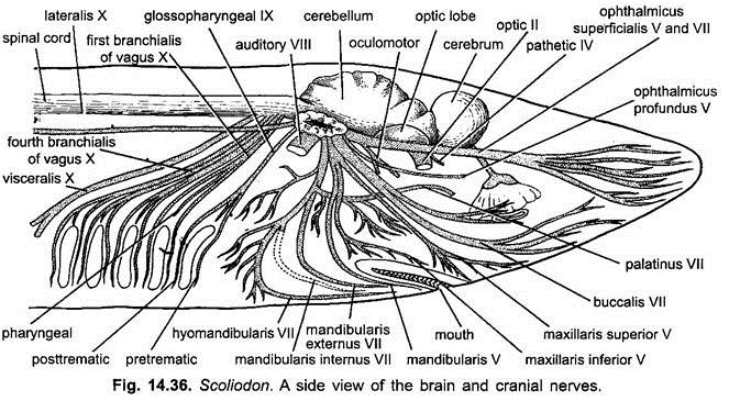
It is a small branch entering the orbit along with the ophthalmicus superficialis of the seventh nerve. It innervates the skin of the snout and is sensory in nature.
(ii) Maxillaris:
It forms two branches, the maxillaris superior and maxillaris inferior. The maxillaris superior is ribbon-like and runs along the floor of the orbit along with buccal branch of VII. It innervates the skin of the upper jaw, while the maxillaris inferior innervates the posterior part of the upper jaw. It is sensory in function.
(iii) Mandibularis:
It runs along the posterior wall of orbit and innervates the muscles of the lower jaw, tongue and gill-region. It is a mixed nerve.
(iv) Ophthalmicus Profundus:
It passes through the inner side of orbit, sends a branch to the eye-ball and goes to the skin of the dorsal surface of the snout. It is sensory in nature.
6. Sixth or Abducens Nerve:
It is a slender nerve arising mid-ventrally from the medulla oblongata. It innervates the posterior rectus muscle of the eye-ball. It is sensory in function.
7. Seventh or Facial Nerve:
It is very large nerve and emerges from the side of medulla oblongata in two bundles- The first bundle is a thick broad ophthalmicus superficialis running forward along the upper border of eye orbit. It along with ophthalmic branch of the fifth nerve goes to the lateral line receptors and ampullae of Lorenzini on the dorsal side of the snout. The second bundle divides into three branches called ramus buccalis, ramus hyomandibularis and ramus palatinus. The facial nerve is a mixed type of nerve having sensory as well as motor fibres.
(i) Ophthalmicus Superficialis:
It innervates the lateral line receptors and ampullae of Lorenzini of the dorsal side of snout.
(ii) Ramus Buccalis:
It runs along the floor of eye orbit and innervates the infra-orbital canal of the snout and its associated groups of ampullae of Lorenzini.
(iii) Ramus Hyomandibularis:
It runs along the posterior wall of orbit and gives out a small branch to the lateral line receptors and then divides into following three branches:
(a) Mandibularis Externus:
It innervates the mandibular canal.
(b) Mandibularis Internus:
It innervates the mucous membrane of the buccal floor.
(c) Hyoidean or Hyomandibular:
It innervates the muscles of the throat (hyoid arch).
(iv) Ramus Palatines:
It runs along the floor of orbit and innervates the roof of the buccal and pharyngeal cavity.
8. Eighth or Auditory Nerve:
It arises close to the origin of fifth and seventh cranial nerves from the side of the medulla and divides immediately into two branches- the vestibular and saccular. It is sensory in function.
(i) Vestibular:
It innervates the membranous labyrinth.
(ii) Saccular:
It innervates the cochlea.
9. Ninth or Glossopharyngeal Nerve:
It arises from the ventro-lateral surface of the medulla behind the origin of sixth nerve, it runs obliquely backwards and divides into three branches- pre-trematic or hyoid branch, post-trematic and pharyngeal. This nerve is mixed in function.
(i) Pre-Trematic:
It runs along the anterior border of the first gill-pouches and supplies the mucous membrane around the first gill-cleft.
(ii) Post-Trematic:
It runs along the posterior border of the first gill-pouches.
(iii) Pharyngeal:
It innervates the mucous membrane of pharynx.
10. Tenth or Vagus Nerve:
Vagus nerve is very large and arises from many roots from the postero-lateral side of medulla oblongata behind the ninth, it divides into three branches- branchialis, visceralis and lateralis.
(i) Branchialis:
Branchialis consists of four main nerves going around the second, third, fourth and fifth gill-pouches. Each nerve consists of a pre-trematic, post-trematic and pharyngeal branches. The branchial nerve-branches supply the mucous membrane around the gill-pouches and pharynx.
(ii) Visceralis:
It is a large nerve which runs backward into the body cavity and innervates the viscera including the alimentary canal, liver, lungs and the heart.
(iii) Lateralis:
Lateralis runs parallel but below the lateral line canal giving branches to the receptors of the lateral line along the length of the body.
An occipital nerve arises behind the vagus and joins the first two spinal nerves to form a hypobranchial nerve which goes to muscles in the floor of the buccal cavity.
The cranial nerves of Scoliodon, as in all fishes, are suited to an aquatic mode of life and are related to the gills, lateral line receptors, and ampullae of Lorenzini, for their innervation the trigeminal, facial, glossopharyngeal, and vagus are specialised.
In land vertebrates the branches of trigeminal and facial which go to the lateral line receptors and various groups of ampullae of Lorenzini have disappeared, as also the branches of the glossopharyngeal and vagus which go to the gills because gills are lost, the lateralis branch of the vagus is also lost with the disappearance of lateral line receptors in terrestrial vertebrates.
B. Spinal Nerves:
The spinal nerves arise in pairs from the sides of the spinal cord at regular intervals along its length. The number of paired spinal nerves is very large, approximately corresponding to the number of vertebrae. Each spinal nerve of each side arises by two roots, a dorsal or sensory root and a ventral or motor root. The ventral root always arises in front of the dorsal root. The dorsal root has a ganglion.
Both the roots run backward for a short distance within the neural canal and perforate the cartilages of the neural arch separately, but unite outside to form a common mixed spinal nerve.
Each spinal nerve divides into three branches:
(i) Ramus dorsalis supplying the skin and muscles of the dorsal body-wall,
(ii) Ramus ventralis supplying the skin and muscles of the ventral body-wall, and
(iii) Ramus communicants containing visceral sensory and motor fibres and joining the autonomic nervous system.
The dorsal root carries somatic sensory, visceral sensory and visceral motor fibres, while the ventral root has somatic motor and visceral motor fibres.
In the region of the pectoral fin spinal nerves three to six join each other near the pectoral girdle to form a cervico-brachial plexus going to the pectoral fin. Spinal nerves seven to eleven pass downwards and send branches to the muscles of ventral body wall and to the pectoral fin.
All spinal nerves from the twelfth to the anus go to body wall muscles. Then nine or ten spinal nerves go to the pelvic fin, some of them joining each other to form a lumbosacral plexus. The remaining spinal nerves go to the caudal region.
III. Autonomic Nervous System:
The autonomic nervous system comprises a paired series of irregularly arranged ganglia situated anteriorly in the dorsal wall of the posterior cardinal sinuses and posteriorly in the dorsal part of the kidney on each side of the mid-dorsal line. The first ganglion is small. The second or gastric ganglion is fairly large, being formed by the fusion of a number of ganglia. It lies immediately behind the post-branchial plexus.
It is connected with numerous fibres from spinal nerves and gives off branches to the viscera. The succeeding ganglia are small. There is always at least one ganglion in each segment, but often there are two or three in a segment. Sometimes the ganglia of successive segments are joined by longitudinal connectives, but there is no definite continuous chain. The posterior ganglia innervate the genital ducts, kidneys, urinary sinus, intestine and rectum.

