In this article we will discuss about the urinogenital organs of dogfish (scoliodon) with the help of suitable diagrams.
In all the vertebrates the excretory and reproductive organs are closely related to each other and are, therefore, dealt together. The two systems together are known as urino- genital system and the organs as urinogenital organs. In Dogfish (Scoliodon), the sexes are separate and the sexual dimorphism is conspicuous. The male Scoliodon possesses two cylindrical hollow copulatory organs, the claspers which are modifications of pelvic fins. The claspers are absent in female.
Male Urinogenital Organs:
1. Excretory Organs:
ADVERTISEMENTS:
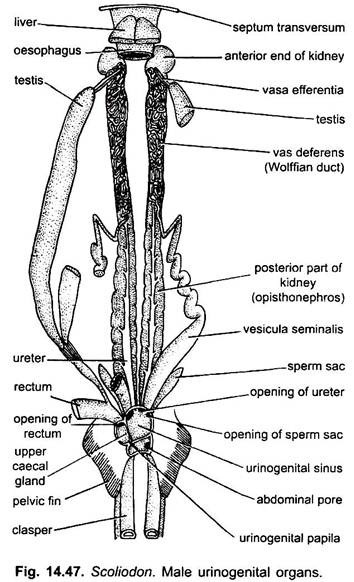
The excretory organs in male consist of pair of kidneys, ureters and unpaired urinogenital sinus. The kidney of Dogfish (Scoliodon) is mesonephric. Each kidney is a long flattened and ribbon-like glandular structure lying dorsally to the peritoneum on either side of the median line and attached to the dorsal abdominal wall. They extend through the entire length of the body cavity from the root of the liver in front up to the cloaca behind.
Each kidney is distinguished into an anterior slender, non-excretory portion, the genital kidney (cranial mesonephros) and a posterior thicker, excretory part, renal kidney (caudal mesonephros). The posterior region of the kidney is greatly thickened, laterally compressed and forms the chief organs of excretion called opisthonephros. While, the anterior region is reduced and becomes comparatively narrow and takes the function of conveying genital products and is, therefore, called the epididymis.
The substance of the opisthonephric kidney is made up of coiled glandular uriniferous tubules. Each uriniferous tubule comprises a Bowman’s capsule enclosing the glomerulus and a coiled renal tubule, several of which open into a common collecting tubule. The renal tubules have a special urea-absorbing segment.
The collecting tubules of each kidney open independently into a common duct, the ureter which finally opens posteriorly into a wide median chamber, the urinogenital sinus. The urinogenital sinus opens into the cloaca through an aperture located at the tip of a small urinogenital papilla.
2. Reproductive Organs:
The reproductive organs include a pair of testes, a pair of vas deferens, and a pair of seminal vesicles.
Testes:
There are two elongated testes lying one on either side of the middle line. Each testis extends from the base of the liver in front to the caecal gland behind. The testes are attached to the dorsal body wall by a fold of peritoneum called the mesorchium and to the caecal gland behind by a non-glandular tissue. Each testis is made up of numerous narrow seminiferous tubules lined with germinal cells which produce sperms by spermatogenesis.
ADVERTISEMENTS:
From each testis arise several thin tubular vasa efferentia which open into the anterior end of a large, narrow closely coiled tube occupying the entire part of the anterior part of the kidney or epididymis but becomes very much enlarged posteriorly to form the wide straight vesicula seminalis or seminal vesicle in which sperms are stored.
The seminal vesicles of both the sides open behind independently into a large triangular chamber, the urinogenital sinus which in its turn opens into the cloaca on an elevated urinogenital papilla. On either side of the urinogenital sinus arises a club-shaped sperm-sac. The function of the sperm-sac is unknown.
Accessory Reproductive Organs:
These are the claspers and siphons. The claspers are grooved elongations of the pelvic fins. The sperms from the cloaca pass into the claspers and are then transferred to the female, thus, acting as intermittent organs in coition. Each clasper has a closed groove with an anterior opening, apopyle lying near the cloaca and a posterior opening called the hypopyle. Sperms enter apopyle from the urinogenital papilla and then transferred to the cloaca of female during coitus.
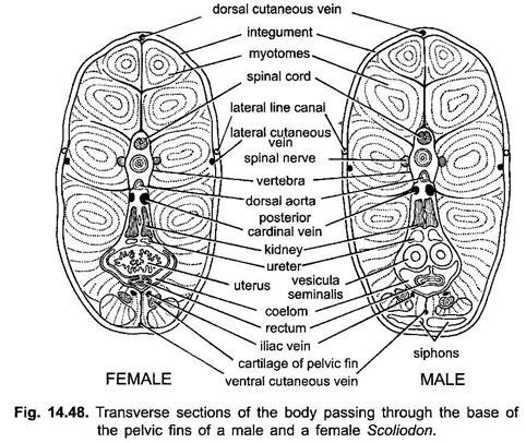
On the ventral side of the body just beneath the skin there is a pair of elongated glandular and muscular sacs, the siphons. These sacs extend anteriorly almost to the level of the posterior margin of the pectoral fine where they end blindly.
Posteriorly these sacs extend as siphon tubes, each of which opens into the anterior opening of clasper of its side. The siphons have no direct communication with the male urinogenital organs except they open into the groove of claspers. It is believed that sea water enters the siphons during copulation and forced down into the clasper groove forcing into the female cloaca.
Female Urinogenital Organs:
1. Excretory Organs:
The excretory organs of female differ from those of the male organs in the following:
(i) There is no direct connection between the kidneys and the genital organs.
ADVERTISEMENTS:
(ii) The anterior part of each kidney is extremely reduced and non-excretory.
(iii) Both the ureters unite behind into a common median ureter which opens by a single median urinary aperture into the triangular chamber, the urinary sinus.
(iv) The urinary sinus receives the ureters only and the genital duct opens directly into the cloaca.
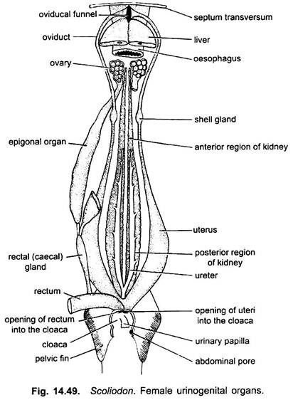
Each kidney of the female also has two parts, an anterior part which is degenerate and reduced to a narrow tapering strand which is non-renal and non-functional, and a broad posterior part which contains masses of coiled uriniferous tubules. From the inner margin of each kidney arises a ureter or Wolffian duct into which open the uriniferous tubules.
The two ureters unite to form a common ureter which opens into a large median urinary sinus. The urinary sinus is formed by the enlarged ends of the Wolffian duct. The urinary sinus opens into the cloaca by an aperture on a urinary papilla.
2. Reproductive Organs:
The female reproductive organs comprise a pair of ovaries, a pair of oviducts, shell gland and a pair of uteri.
Ovaries:
The ovaries are paired organs varying in form and size according to the age of Dogfish (Scoliodon). The lobulated overies are situated, one on either side of the vertebral column, just behind the base of liver. They are suspended in the body cavity, from the dorsal abdominal wall by a fold of peritoneum, the mesovarium. Round follicles project from each ovary, each follicle contains an ovum. Between the ovary in front and caecal gland behind extends a long tubular strand of tissue, the epigonal organ.
Oviducts, the two oviducts are the well-developed Mullerian ducts extending the entire length of the body cavity. Each oviduct is a long thick-walled muscular tube and their anterior narrow ends unite mid-ventrally beneath the oesophagus near septum transversum and opens into the coelom by a single longitudinal slit, called the oviducal funnel or ostium.
Each oviduct runs backward for a short distance and then enlarges to form the shell gland or oviducal gland. It secretes a thin membrane over the descending fertilised eggs. The oviduct then again narrows and then it again dilates to form a sac-like uterus.
The uteri of both the sides unite to form a short vagina which opens into the cloaca by a large aperture. A fold of mucous membrane of this region separates the vagina from the cloaca, and acts as a valve which closes the vagino- cloacal aperture during the development of the embryo within the uterus.
The oviducts have no direct communication with the ovaries so that mature ova are at first shed into the abdominal cavity and later forced by the action of the body-muscles into the oviducts through the single oviducal funnel. Fertilisation occurs in the section of oviduct between the oviducal funnel and the shell-gland. The fertilised ova come into the uterus.


