Development of Herdmania has been described by Sebastian (1953) and Das (1957). Development is indirect involving a free swimming tadpole larva. Herdmania (a tunicate) is the most appropriate one of the best examples of retrogressive metamorphosis in which the highly developed tadpole larva undergoes retrogressive changes to become the most degenerated and sedentary adult. The phenomenon of retrogressive metamorphosis is peculiar to urochordates and provides a clue to their chordate nature.
Gametes:
Spermatozoa:
The sperm of Herdmania is flagellated and about 4 μm in length. Each sperm has an anterior head bearing a middle nucleus and capped with a beak-like acrosome, a middle piece with neck and a very long tail. According to Das (1936), sperms of Herdmania are polymorphic, i.e., three types of sperms having acrosome either shorter, equal or longer than the head.
Ova:
ADVERTISEMENTS:
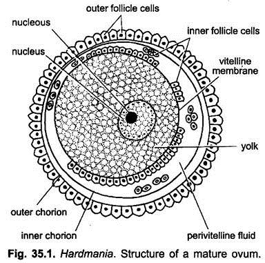
The ova or unfertilised eggs are microlecithal having distinct animal and vegetal poles. Mature ovum is about 0.3 mm in diameter containing an eccentric nucleus with a prominent nucleolus. The egg is surrounded by an inner vitelline membrane secreted by the ovum which is closely applied to it, and outer to it are two chorion membranes inner and outer secreted by follicular cells of the ovary.
The space between inner chorion and vitelline membrane is the perivitelline space filled with perivitelline fluid. The perivitelline space contains a large number of small-sized inner follicle cells or test cells, some of which float in the fluid and most of them are attached to vitelline membrane.
These cells nourish the ovum and secrete a chorion digesting enzyme to facilitate hatching. Follicle cells are also found attached to the outer surface of chorion membrane. These cells keep the ovum floating in outer perivitelline fluid. Maturation of ovum occurs in sea water.
Fertilisation:
ADVERTISEMENTS:
Fertilisation is external and cross-fertilisation. Spermatozoon penetrates the ovum nearer to vegetal pole. Polyspermy is not allowed by the chorion layers. After fertilisation, various cytoplasmic regions are rearranged by a process known as ooplasmic segregation, i.e., yellow and clear cytoplasm segregates in the lower hemisphere with a cap of clear cytoplasm over it and gray yolk-laden endoplasm occupies the upper two-thirds of the egg.
Presumptive Areas:
The entry of sperm into ovum results in movements of ovum’s cytoplasm (ooplasm), so that the fertilised egg or zygote is mosaic, i.e, it shows presumptive areas with predictable future.
Embryonic or Prelarval Development:
Cleavage:
ADVERTISEMENTS:
Cleavage is determinate. The zygote undergoes holoblastic, somewhat unequal and determinate cleavage, i.e., the fate of blastomeres is predetermined. The first cleavage is vertical, meridional and divides the gray crescent into two unequal parts. Second cleavage is also vertical but at right angle to the first. The resultant four blastomeres are different. Two smaller blastomeres are at posterior side and two larger blastomeres are at front. Only two of these four contain parts of gray crescent.
The third cleavage is horizontal and passes little above the equator which divides the blastomeres into 8 cells arranged in two tiers. Fourth cleavage results into 16 blastomeres. Subsequently, cleavage becomes irregular and finally results into a solid ball of cells called morula. A single-layered flat coeloblastula with an internal fluid-filled segmentation cavity or blastocoel is formed at 64-cell stage. In all it takes about 110 minutes.
Thus, a round 64-celled compact blastula is formed having a blastocoel in the interior. The upper smaller cells are micromeres or ectoderm, and lower larger cells are macromeres or endoderm.
Gastrulation:
It takes place by emboly or invagination of macromeres, which now enclose an archenteron. The wide ventral opening of the archenteron is the blastopore, the archenteron increases obliterating the blastocoel.
On the completion of gastrulation, the blastopore closes and the embryo begins to lengthen along the embryonic axis. The upper surface or neural plate of the elongating gastrula invaginates along the mid-line to form a groove which subsequently forms the neural tube. This stage of the embryo is neurala.
The anterior portion of neural tube remains wider and develops into brain vesicle, and the posterior narrow portion of neural tube forms the spinal cord. For a short time, the brain- vesicle communicates to the exterior through neuropore.
The hinder part of neurula becomes extended into a tail, into which neural tube, notochord, mesoderm and mesenchyme are extended. With further development, the neurula develops into an ascidian tadpole larva, which ruptures the egg membranes and becomes free-swimming.
Larval Development:
ADVERTISEMENTS:
The blastopore closes and develops a rudiment of tail. The embryo elongates and forms a tailed larva. The presumptive notochordatal cells separated from the roof of the archenteron,occupy the central core of larval tail.
Archenteron produces presumptive mesoderm as solid bands and not as hollow coelomic sacs as in Balanoglossus or Branchiostoma. About 8 hours after fertilisation, the chorions rupture, probably dissolved by an enzyme secreted by the inner follicle cells, and the fully formed larva hatches out to become free-swimming.
Tadpole Larva:
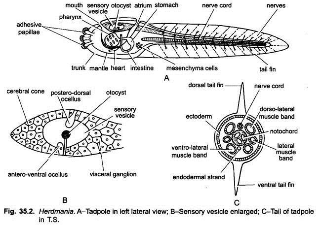
The fully developed ascidian tadpole is minute, about 1.5 mm long and motile. Its body is covered by a thin tunic or test secreted by the ectoderm. It has an anterior short oval head or trunk and a posterior long tail.
1. Head or Trunk:
At the anterior end of the trunk are three adhesive papillae made of ectodermal cells. The nervous system consists of an anterior enlarged hollow sense vesicle or brain vesicle. The sense vesicle is continued posteriorly into a solid, thick mass of nerve cells, the visceral ganglion, it is continued posteriorly into a nerve cord which is hollow and continued into the tail. It lies mid-dorsally.
The sense vesicle contains a pigmented statocyst (otocyst) and two pigmented unequal ocelli or simple eyes as sense organs.
The alimentary canal begins from antero-dorsal mouth leading into narrow branchial siphon, a large sac-like pharynx, short oesophagus, stomach, intestine and a small rectum. The anus opens into the left side of atrium. The pharynx has a ventral endostyle and also a pair of large gill-slits or stigmata.
Each gill-slit finally splits to form six stigmata on either lateral side An atrial cavity is formed around the pharynx laterally and dorsally and opens to the exterior by an atrial aperture lying dorsally. The heart and pericardium is located postero-ventrally in between pharynx and stomach. Mesenchyme cells found scattered all over the body beneath the ectoderm and in a mass in the posterior region of trunk.
2. Tail:
It is a powerful locomotory organ being laterally compressed and pointed terminally. Tail is about 0.9 mm long and is fringed with a vertical continuous tail fin formed by the test and marked with oblique striations looking like fin rays. It contains a dorsal tubular nerve cord, a notochord beneath the nerve cord and lateral muscle bands. The anterior end of notochord slightly projects into the trunk region.
The ascidian tadpole larva is unquestionably a true chordate, having a dorsal hollow nerve tube, a notochord in the tail and paired gill-slits in the pharynx and after about three hours of active, non-feeding and free-swimming existence, it sinks to the bottom.
Habits and Habitat of Larva:
Just after hatching, the larva is photopositive and geonegative. It cannot feed because its mouth is still closed by test. After a short active free-swimming existence lasting about 3 to 4 hours, the larva becomes geopositive, photonegative and sluggish.
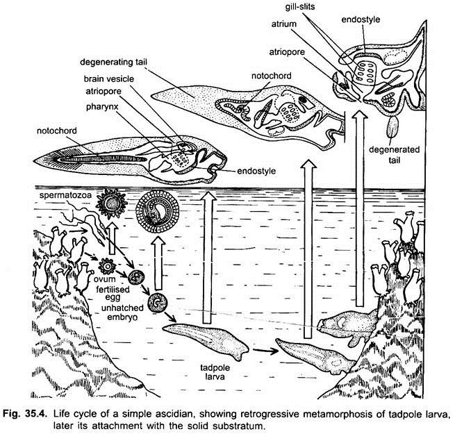
It sinks to the bottom, attaches itself upside down to a suitable hard substratum by adhesive papillae and undergoes rapid degeneration or retrogressive metamorphosis to attain adulthood. According to Berril (1955) the selection of a suitable habitat is essential as the larva may not survive on any other habitat or may get suffocated by the bottom mud and detritus.
Retrogressive Metamorphosis:
Metamorphosis Gr., meta = after + morphe = form + osis = state) is the shape change in form during post-embryonic development and in many cases, signals a dramatic change in habitat of the animal such as pelagic to benthic existence.
Metamorphosis of the ascidian larva is unique and begins almost explosively. It involves transformation of an active non-feeding, pelagic, lecithotrophic (i.e., that feeds on its own yolk reserves) and tailed larva having many advanced features such as axial notochord, dorsal neural tube and special sense organs, into an inert, sedentary or sessile, simple (primitive) and plankotrophic filter feeding adult with only a phaynx with stigmata and endostyle, both indicating the chordate features of adult ascidian.
This type of metamorphosis which shows degenerative or retrogressive changes from larva to adult is called retrogressive metamorphosis.
It involves the following three types of changes:
(i) Retrogressive,
(ii) Progressive and
(iii) Molecular changes.
(i) Retrogressive Changes:
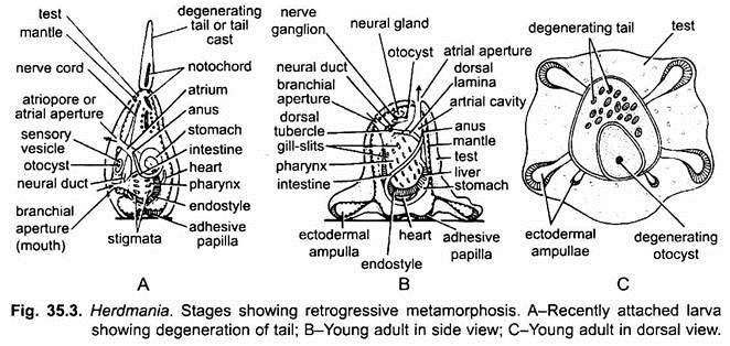
These changes involve destruction and disappearance of some of the larval structures such as follows:
a. Long tail of larva with caudal fin shortens and finally disappears.
b. Caudal muscles, nerve cord and notochord disappear as they break down and are consumed by phagocytes.
c. Larval sense organs (the ocellus and the otolith) are lost and sensory vesicle is transformed into an adult cerebral ganglion.
d. Adhesive papillae disappear completely.
e. Anterior region between point of attachment (adhesive papillae) and mouth shows rapid growth, while original dorsal side with atriopore stops growth. This causes shifting of mouth through 90°. Therefore, the final branchial and atrial apertures in the adult represent the original anterior and dorsal sides of the larva.
(ii) Progressive Changes:
Some larval structures necessary for survival become more elaborated and specialised in each adult, such as:
a. Due to loss of tail, the trunk becomes pear-shaped and four larger ectodermal ampullae grow out of its four corners. These ampullae firmly anchor the metamorphosing tadpole to the substratum and also serve for respiration as a blood-like fluid keeps circulating through them. Soon two more smaller ectodermal ampullae appear dorso-laterally.
b. Anterior region between point of attachment (adhesive papillae) and mouth exhibits rapid growth, while original dorsal side with atriopore stops growth. This causes shifting of mouth through 90°. The body too rotates so that general form of the adult sessile organism is assumed.
c. Adult neural glands and nerve or cerebral ganglion are formed by the neural tube and trunk ganglion come to lie mid-dorsally between mouth and atriopore. The trunk ganglion itself persists as visceral nerve.
d. With the absorption of its test covering, the mouth becomes functional and filter-mode of feeding by incoming ciliary water currents.
e. Pharynx greatly enlarges, develops blood vessels and stigmata multiply rapidly, forming the branchial sac.
f. Stomach enlarges, intestine elongates and gets curved and liver develops.
g. Atrial cavity becomes more extensive.
h. Circulatory system with heart and pericardium develops.
i. Gonads and gonoducts develop from larval mesodermal cells.
j. Test or tunic spreads to cover entire animal, becomes thick, tough and vascular and attaches the animal by forming a foot if necessary.
Thus, foregoing metamorphic changes mark the beginning of a sedentary, actively feeding, sexual adult life which soon starts producing gametes, first ova and later sperms.
(iii) Molecular Changes:
Manket and Cowden (1965) studied the molecular changes which take place during metamorphosis. They studied the metabolism of protein and nucleic acid and pointed out that some protein synthesis occurs throughout the development but with the outset of metamorphosis, extensive degradation of proteins begins followed by rapid synthesis of new proteins.
Embryological Significance of Ascidian Tadpole:
The presence of a tadpole larva in the life history of Herdmania and other ascidians is significant in the following ways:
a. Taxonomic Significance:
The tadpole larva possesses true chordate characters such as notochord and dorsal tubular nerve cord, which are lacking in the adult. Thus, the ascidian larva provides the clue for including the ascidian under the phylum Chordata. Without tadpole larva, the true nature and taxonomic position of degenerate sedentary adult ascidians would have remained uncertain.
b. Phylogenetic Significance:
On the basis of recapitulation, the ascidian larva possessing the chordate features is considered as the relic of the free-swimming ancestral vertebrates.
c. Dispersal:
The adult ascidians being sedentary, the free-swimming habit of the larva provides the only means of dispersal of the species. It also provides chances of selecting better sites regarding food and protection, thus, ensuring survival of the race.
d. Embryological Significance:
Ascidians provide best example of mosaic eggs with a well- organised, pre-patterned and well differentiated ooplasm and highly determinate type of development. Moreover, ascidians are the only chordates exhibiting true retrogressive metamorphosis.
The egg cortex in case of ascidians is the site of morphogenetic patterning related to polar, bilateral and general organisation of developing egg. Besides this, cleavage in ascidians tends to segregate cytoplasmic territories, having different biological, histochemical and biochemical properties.


